Tuesday Poster Session
Category: Stomach
P5126 - E. histolytica Gastric Infection Superadded to Gastric Adenocarcinoma: An Uncommon Case Report
Tuesday, October 29, 2024
10:30 AM - 4:00 PM ET
Location: Exhibit Hall E

Has Audio
- MG
Monica C. Guillen
Universidad Católica de Santiago de Guayaquil
Guayaquil, Guayas, Ecuador
Presenting Author(s)
Francisco Cano, MD1, Monica C. Guillen, 2, Sergio A. Manzur, 2, Wilton J. Cueva, 2, Pablo S. Bermeo, 3
1Hospital Alfredo G. Paulson, Guayaquil, Guayas, Ecuador; 2Universidad Católica de Santiago de Guayaquil, Guayaquil, Guayas, Ecuador; 3Universidad Católica de Santiago de Guayaquil, Daule, Guayas, Ecuador
Introduction: Entamoeba histolytica, a protozoan parasite primarily implicated in amebiasis, predominantly invades the colon and occasionally the liver, resulting in dysentery and liver abscesses. Nonetheless, its invasion of other tissues and organs is uncommon and not well-documented, physicians should consider this phenomenon and the potential coexistence with malignant lesions in patients from endemic regions.
Case Description/Methods: A 38-year-old man from Ecuador presented with three episodes of melena accompanied by fever, hyporexia, asthenia and substantial weight loss (33lbs). He has no significant medical background, and a diagnosis of gastrointestinal hemorrhage was established.
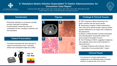
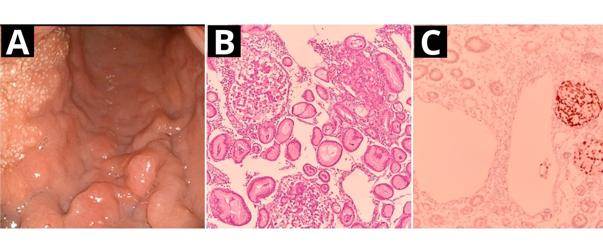
A high-resolution CT scan (HRCT) depicted a diffuse thickening of the gastric chamber wall and severe ascites. Subsequently, a gastroscopy was performed, where a friable erythematous lesion with poorly defined borders was observed, featuring whitish papules distributed on its surface with a strawberry-like appearance. Its biopsy unveiled abundant rounded trophozoites with intracytoplasmic eosinophilic bodies in the lamina propia of the gastric mucosa. The immunohistochemistry (IH) findings of CDX2+ suggested malignancy with neoplastic cells of the GI tract indicative of adenocarcinoma. With the proven diagnosis, the patient was referred to the oncology service and began treatment with 5-fluorouracil. During the ensuing weeks, there was no observable amelioration in the patient's condition and succumbed after a treatment duration of 7 weeks.
Discussion: An exhaustive literature search was conducted in multiple databases, revealing no previously reported cases. Although the gastric tissue is not a common site for Entamoeba histolytica colonization, the presence of adenocarcinoma in this instance may have created favorable conditions for such colonization. Therefore, this case emphasizes the importance of considering gastrointestinal malignancies, in the differential diagnosis of parasitic infections of atypical sites of the digestive tract.

Disclosures:
Francisco Cano, MD1, Monica C. Guillen, 2, Sergio A. Manzur, 2, Wilton J. Cueva, 2, Pablo S. Bermeo, 3. P5126 - E. histolytica Gastric Infection Superadded to Gastric Adenocarcinoma: An Uncommon Case Report, ACG 2024 Annual Scientific Meeting Abstracts. Philadelphia, PA: American College of Gastroenterology.
1Hospital Alfredo G. Paulson, Guayaquil, Guayas, Ecuador; 2Universidad Católica de Santiago de Guayaquil, Guayaquil, Guayas, Ecuador; 3Universidad Católica de Santiago de Guayaquil, Daule, Guayas, Ecuador
Introduction: Entamoeba histolytica, a protozoan parasite primarily implicated in amebiasis, predominantly invades the colon and occasionally the liver, resulting in dysentery and liver abscesses. Nonetheless, its invasion of other tissues and organs is uncommon and not well-documented, physicians should consider this phenomenon and the potential coexistence with malignant lesions in patients from endemic regions.
Case Description/Methods: A 38-year-old man from Ecuador presented with three episodes of melena accompanied by fever, hyporexia, asthenia and substantial weight loss (33lbs). He has no significant medical background, and a diagnosis of gastrointestinal hemorrhage was established.
A high-resolution CT scan (HRCT) depicted a diffuse thickening of the gastric chamber wall and severe ascites. Subsequently, a gastroscopy was performed, where a friable erythematous lesion with poorly defined borders was observed, featuring whitish papules distributed on its surface with a strawberry-like appearance. Its biopsy unveiled abundant rounded trophozoites with intracytoplasmic eosinophilic bodies in the lamina propia of the gastric mucosa. The immunohistochemistry (IH) findings of CDX2+ suggested malignancy with neoplastic cells of the GI tract indicative of adenocarcinoma. With the proven diagnosis, the patient was referred to the oncology service and began treatment with 5-fluorouracil. During the ensuing weeks, there was no observable amelioration in the patient's condition and succumbed after a treatment duration of 7 weeks.
Discussion: An exhaustive literature search was conducted in multiple databases, revealing no previously reported cases. Although the gastric tissue is not a common site for Entamoeba histolytica colonization, the presence of adenocarcinoma in this instance may have created favorable conditions for such colonization. Therefore, this case emphasizes the importance of considering gastrointestinal malignancies, in the differential diagnosis of parasitic infections of atypical sites of the digestive tract.

Figure: (A) Gastroscopy of gastric body showing a friable erythematous lesion with poorly defined borders, featuring whitish papules distributed on its surface. (B) Biopsy evidence histological section with H&E showing superficial erosion with dense lymphoplasmacytic inflammatory infiltrate of lamina propria, dilatations occupied by abundant rounded trophozoites with intracytoplasmic eosinophilic bodies some of them with erythrocyte ingestion in the lamina propria of the gastric mucosa, preserving the glandular architecture. (C) Immunohistochemical profile (CDX2+), agrees with the histopathological findings and indicates the presence of malignant and metastatic neoplastic cells of the GI tract for adenocarcinomas.
Disclosures:
Francisco Cano indicated no relevant financial relationships.
Monica Guillen indicated no relevant financial relationships.
Sergio Manzur indicated no relevant financial relationships.
Wilton Cueva indicated no relevant financial relationships.
Pablo Bermeo indicated no relevant financial relationships.
Francisco Cano, MD1, Monica C. Guillen, 2, Sergio A. Manzur, 2, Wilton J. Cueva, 2, Pablo S. Bermeo, 3. P5126 - E. histolytica Gastric Infection Superadded to Gastric Adenocarcinoma: An Uncommon Case Report, ACG 2024 Annual Scientific Meeting Abstracts. Philadelphia, PA: American College of Gastroenterology.

