Tuesday Poster Session
Category: Stomach
P5128 - Gastric Diverticulitis Imitating an Ulcerated Gastric Neoplasia
Tuesday, October 29, 2024
10:30 AM - 4:00 PM ET
Location: Exhibit Hall E

Has Audio

Matthew M. Eganhouse, MD
Rush University Medical Center
Chicago, IL
Presenting Author(s)
Matthew M. Eganhouse, MD, Kanika Garg, MD, Ajaypal Singh, MD, Michael Brown, MD, MACM
Rush University Medical Center, Chicago, IL
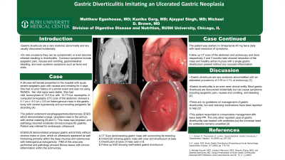
Introduction: Gastric diverticula are a rare anatomic abnormality and are usually discovered incidentally. We present a case of a patient with symptomatic gastric diverticulitis masquerading as a neoplastic lesion. Although initial work up was concerning for malignancy, biopsies and endoscopic ultrasound (EUS) findings were consistent with inflammatory changes, and patient had resolution of symptoms with conservative management. This case demonstrates that gastric diverticulitis, while rare, should be on the differential for patients with vague abdominal symptoms.
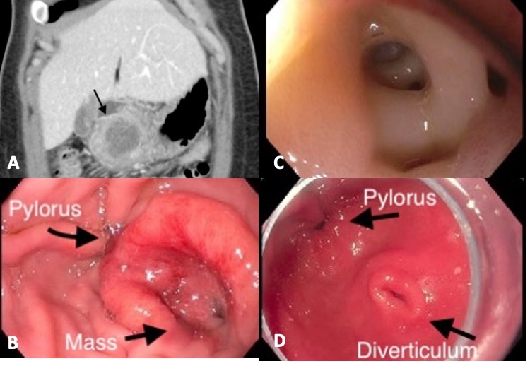
Case Description/Methods: A 29-year-old female presented with 4 days of epigastric pain with nausea and vomiting. Her vital signs were stable. Labs were remarkable for leukocytosis of 13.5 KuL. A computed tomography (CT) scan showed a heterogenous mass in the gastric body with central hypodensity and surrounding perigastric fat stranding. She underwent esophagogastroduodenoscopy (EGD) which demonstrated a large, ulcerated mass in the antrum with central cratering. Biopsies of this mass returned as moderate chronic/nonspecific gastritis. Subsequent EGD/EUS demonstrated enlarged gastric antral folds with wall thickening primarily within the deep mucosa and submucosa with heterogenous echogenicity. Fine needle biopsies showed fibrous tissue with chronic inflammation within the submucosa. She was started on twice daily proton pump inhibitor (PPI) with rapid resolution of symptoms. Follow up CT scan and endoscopy 3 months later showed resolution of the mass and healthy antral mucosa with a single gastric diverticulum present without any surrounding inflammation.
Discussion: Gastric diverticula are rare anatomic abnormalities with an estimated prevalence of 0.05 to 0.1% at endoscopy. Gastric diverticulitis is an even rarer clinical entity. Most gastric diverticula are discovered incidentally but can cause symptoms including epigastric pain, nausea and vomiting, and bleeding. There are no current guidelines on management of gastric diverticulitis, but acid reducing medications have been reported to help. In this case, a gastric diverticulum became infected resulting in luminal thickening, inflammatory changes, and obstructive symptoms mimicking neoplasia. The short onset of symptoms however was more consistent with an acute inflammatory process. This patient responded to conservative management with twice daily PPI. The only other reported case of gastric diverticulitis was treated with antibiotics but the universal need for antibiotics remains unsettled.

Disclosures:
Matthew M. Eganhouse, MD, Kanika Garg, MD, Ajaypal Singh, MD, Michael Brown, MD, MACM. P5128 - Gastric Diverticulitis Imitating an Ulcerated Gastric Neoplasia, ACG 2024 Annual Scientific Meeting Abstracts. Philadelphia, PA: American College of Gastroenterology.
Rush University Medical Center, Chicago, IL
Introduction: Gastric diverticula are a rare anatomic abnormality and are usually discovered incidentally. We present a case of a patient with symptomatic gastric diverticulitis masquerading as a neoplastic lesion. Although initial work up was concerning for malignancy, biopsies and endoscopic ultrasound (EUS) findings were consistent with inflammatory changes, and patient had resolution of symptoms with conservative management. This case demonstrates that gastric diverticulitis, while rare, should be on the differential for patients with vague abdominal symptoms.
Case Description/Methods: A 29-year-old female presented with 4 days of epigastric pain with nausea and vomiting. Her vital signs were stable. Labs were remarkable for leukocytosis of 13.5 KuL. A computed tomography (CT) scan showed a heterogenous mass in the gastric body with central hypodensity and surrounding perigastric fat stranding. She underwent esophagogastroduodenoscopy (EGD) which demonstrated a large, ulcerated mass in the antrum with central cratering. Biopsies of this mass returned as moderate chronic/nonspecific gastritis. Subsequent EGD/EUS demonstrated enlarged gastric antral folds with wall thickening primarily within the deep mucosa and submucosa with heterogenous echogenicity. Fine needle biopsies showed fibrous tissue with chronic inflammation within the submucosa. She was started on twice daily proton pump inhibitor (PPI) with rapid resolution of symptoms. Follow up CT scan and endoscopy 3 months later showed resolution of the mass and healthy antral mucosa with a single gastric diverticulum present without any surrounding inflammation.
Discussion: Gastric diverticula are rare anatomic abnormalities with an estimated prevalence of 0.05 to 0.1% at endoscopy. Gastric diverticulitis is an even rarer clinical entity. Most gastric diverticula are discovered incidentally but can cause symptoms including epigastric pain, nausea and vomiting, and bleeding. There are no current guidelines on management of gastric diverticulitis, but acid reducing medications have been reported to help. In this case, a gastric diverticulum became infected resulting in luminal thickening, inflammatory changes, and obstructive symptoms mimicking neoplasia. The short onset of symptoms however was more consistent with an acute inflammatory process. This patient responded to conservative management with twice daily PPI. The only other reported case of gastric diverticulitis was treated with antibiotics but the universal need for antibiotics remains unsettled.

Figure: A. CT Scan with gastric mass B. Diverticula inside inflamed gastric diverticulum C. Initial EGD with inflamed gastric diverticulum D. 3 month follow up EGD with healed antral gastric diverticulum
Disclosures:
Matthew Eganhouse indicated no relevant financial relationships.
Kanika Garg indicated no relevant financial relationships.
Ajaypal Singh indicated no relevant financial relationships.
Michael Brown indicated no relevant financial relationships.
Matthew M. Eganhouse, MD, Kanika Garg, MD, Ajaypal Singh, MD, Michael Brown, MD, MACM. P5128 - Gastric Diverticulitis Imitating an Ulcerated Gastric Neoplasia, ACG 2024 Annual Scientific Meeting Abstracts. Philadelphia, PA: American College of Gastroenterology.
