Tuesday Poster Session
Category: Stomach
P5138 - A Rare Case of Malignant Melanoma With Gastric Metastases
Tuesday, October 29, 2024
10:30 AM - 4:00 PM ET
Location: Exhibit Hall E

Has Audio
- JJ
Jonathan Jeong, DO
Lenox Hill Hospital, Northwell Health
New York, NY
Presenting Author(s)
Jonathan Jeong, DO1, Jeffrey Dong, MD2
1Lenox Hill Hospital, Northwell Health, New York, NY; 2Lenox Hill Hospital, Northwell Health, Long Island City, NY
Introduction: Malignant melanoma (MM) with gastric metastases is a rare occurrence accounting for only an estimated 20% of all gastrointestinal (GI) tract involvement. Although malignant melanoma remains the most common carcinoma to metastasize to the GI tract, the incidence of symptomatic GI involvement is 1-5% of MM patients. We present a case of gastric metastases arising from malignant melanoma to raise awareness of this uncommon association.
Case Description/Methods: An 80-year-old female with End-stage renal disease on hemodialysis with multiple arteriovenous fistulas, and a remote history of acral melanoma of the left foot presented with purulent drainage from a right forearm arteriovenous graft along with blood cultures from a recent admission revealing E. Faecalis bacteremia. Initial laboratory workup was notable for anemia, with hemoglobin 8.2 gm/dL and hematocrit 26.3%. The basic metabolic panel, liver function tests, and coagulation profile were largely unremarkable.
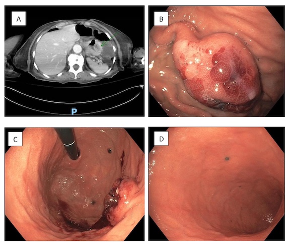
During the hospital course, the patient underwent a complete graft excision and was continued on intravenous antibiotics. As the patient continued to have persistent fevers along with melena in the setting of bacteremia, a computed tomography (CT) scan of the abdomen with contrast was performed which revealed focal mural hypo enhancement and probable ulceration in the gastric fundus. An upper endoscopy was performed, and findings included a mass with central ulceration in the upper stomach body, black-colored nodules in the stomach fundus and lower body, and a Mallory Weiss tear in the cardia. Multiple cold biopsies were obtained, and pathology revealed an immunohistochemical staining profile positive for HMB45, Melan-A, and S100 consistent with malignant melanoma. The patient decided to undergo single-agent immunotherapy with pembrolizumab but was found to have progression of disease after three cycles and was subsequently switched to imatinib for therapy.
Discussion: Malignant melanoma is well known for its ability to metastasize to virtually any organ, but gastric localization is highly unusual and not well reported due to its non-specific symptoms resulting in a paucity of radiologic and endoscopic workup. In our case, the patient initially presented with bacteremia with recurrent fevers, melena and obtained a CT scan and subsequent endoscopy which ultimately led to the diagnosis. The rare association of malignant melanoma with gastric metastases remains important to recognize for prompt diagnosis and treatment initiation.

Disclosures:
Jonathan Jeong, DO1, Jeffrey Dong, MD2. P5138 - A Rare Case of Malignant Melanoma With Gastric Metastases, ACG 2024 Annual Scientific Meeting Abstracts. Philadelphia, PA: American College of Gastroenterology.
1Lenox Hill Hospital, Northwell Health, New York, NY; 2Lenox Hill Hospital, Northwell Health, Long Island City, NY
Introduction: Malignant melanoma (MM) with gastric metastases is a rare occurrence accounting for only an estimated 20% of all gastrointestinal (GI) tract involvement. Although malignant melanoma remains the most common carcinoma to metastasize to the GI tract, the incidence of symptomatic GI involvement is 1-5% of MM patients. We present a case of gastric metastases arising from malignant melanoma to raise awareness of this uncommon association.
Case Description/Methods: An 80-year-old female with End-stage renal disease on hemodialysis with multiple arteriovenous fistulas, and a remote history of acral melanoma of the left foot presented with purulent drainage from a right forearm arteriovenous graft along with blood cultures from a recent admission revealing E. Faecalis bacteremia. Initial laboratory workup was notable for anemia, with hemoglobin 8.2 gm/dL and hematocrit 26.3%. The basic metabolic panel, liver function tests, and coagulation profile were largely unremarkable.
During the hospital course, the patient underwent a complete graft excision and was continued on intravenous antibiotics. As the patient continued to have persistent fevers along with melena in the setting of bacteremia, a computed tomography (CT) scan of the abdomen with contrast was performed which revealed focal mural hypo enhancement and probable ulceration in the gastric fundus. An upper endoscopy was performed, and findings included a mass with central ulceration in the upper stomach body, black-colored nodules in the stomach fundus and lower body, and a Mallory Weiss tear in the cardia. Multiple cold biopsies were obtained, and pathology revealed an immunohistochemical staining profile positive for HMB45, Melan-A, and S100 consistent with malignant melanoma. The patient decided to undergo single-agent immunotherapy with pembrolizumab but was found to have progression of disease after three cycles and was subsequently switched to imatinib for therapy.
Discussion: Malignant melanoma is well known for its ability to metastasize to virtually any organ, but gastric localization is highly unusual and not well reported due to its non-specific symptoms resulting in a paucity of radiologic and endoscopic workup. In our case, the patient initially presented with bacteremia with recurrent fevers, melena and obtained a CT scan and subsequent endoscopy which ultimately led to the diagnosis. The rare association of malignant melanoma with gastric metastases remains important to recognize for prompt diagnosis and treatment initiation.

Figure: A. Gastric fundus focal mural hypo-enhancement on CT scan
B. Non-bleeding 2cm submucosal mass with patchy erythema and central cratered ulceration in proximal stomach body along the greater curvature.
C. Black-colored lesions ranging in size from 2mm to 4mm in the stomach fundus
D. Black-colored lesion in the distal stomach body
B. Non-bleeding 2cm submucosal mass with patchy erythema and central cratered ulceration in proximal stomach body along the greater curvature.
C. Black-colored lesions ranging in size from 2mm to 4mm in the stomach fundus
D. Black-colored lesion in the distal stomach body
Disclosures:
Jonathan Jeong indicated no relevant financial relationships.
Jeffrey Dong indicated no relevant financial relationships.
Jonathan Jeong, DO1, Jeffrey Dong, MD2. P5138 - A Rare Case of Malignant Melanoma With Gastric Metastases, ACG 2024 Annual Scientific Meeting Abstracts. Philadelphia, PA: American College of Gastroenterology.
