Tuesday Poster Session
Category: Stomach
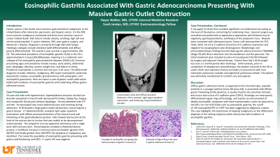
P5085 - Eosinophilic Gastritis Associated With Gastric Adenocarcinoma Presenting With Massive Gastric Outlet Obstruction
Tuesday, October 29, 2024
10:30 AM - 4:00 PM ET
Location: Exhibit Hall E

Has Audio

Hayes M. Walker, MD
University of Tennessee Health Science Center
Memphis, TN
Presenting Author(s)
Hayes Walker, MD, Ronald Scott. Jordan, MD
University of Tennessee Health Science Center, Memphis, TN
Introduction: Gastric cancer is the fifth most common malignancy worldwide and third most common cause of cancer death. The overall 5-year survival is approximately 10%. Eosinophilic gastritis prevalence in the US is about 6.3/100,000 people, making it a rare subtype of eosinophilic gastrointestinal diseases. Peripheral eosinophilia is common but not seen in all cases. Here we report a case of gastric outlet obstruction with evidence of eosinophilic gastritis and underlying diffuse gastric adenocarcinoma.
Case Description/Methods: A 54-year-old male presented to the ED with decreased PO intake and fatigue. He was admitted for UTI and AKI. Abdominal pain developed during admission. Imaging revealed large gastric outlet obstruction, RUQ malrotation, diffuse proximal distention of the stomach and wall thickening of the gastroduodenal junction. Initial concern for gastric bezoar. EGD showed obstruction at the antrum due to torsion and was unable to be decompressed. Biopsies revealed a thickened lamina propria and multifocal increase in eosinophils ( >30/HPF and focally >100/HPF). A diagnosis of eosinophilic gastritis was established. Exploratory laparotomy was performed with bilateral truncal vagotomy, gastrojejunostomy, and biopsy of prepyloric mass. Biopsies were consistent with adenocarcinoma. Tumor cells were positive for CK7, CK20, CDX2, CA 19–9, E-cadherin (focal loss of E-cadherin expression). The patient was diagnosed with gastric adenocarcinoma T4bN0M0 (Stage III) with direct extension into the duodenum and the pancreas. Treatment plan was to include neoadjuvant FOLFOX followed by surgery and adjuvant chemo. Prior to completion of neoadjuvant chemo, the patient returned to the ED in septic shock and respiratory failure secondary to pneumonia with new metastatic pulmonary nodules and segmental pulmonary emboli. The patient was ultimately transitioned to comfort care and expired.
Discussion: Diffuse gastric adenocarcinoma typically presents in a younger patient, is usually found in the proximal stomach, and occurs due to loss of the E-cadherin gene (CDH1). Histology can show poorly differentiated signet ring cells. Reports are documented of deeply eosinophilic cytoplasm with bizarre pleomorphic nuclei. The cutoff of >30 eosinophils/HPF in 5 HPF has been suggested as diagnostic criteria for eosinophilic gastritis. This case highlights the importance of keeping malignancy on the differential diagnosis in the setting of gastric outlet obstruction with evidence of eosinophilic gastritis.

Disclosures:
Hayes Walker, MD, Ronald Scott. Jordan, MD. P5085 - Eosinophilic Gastritis Associated With Gastric Adenocarcinoma Presenting With Massive Gastric Outlet Obstruction, ACG 2024 Annual Scientific Meeting Abstracts. Philadelphia, PA: American College of Gastroenterology.
University of Tennessee Health Science Center, Memphis, TN
Introduction: Gastric cancer is the fifth most common malignancy worldwide and third most common cause of cancer death. The overall 5-year survival is approximately 10%. Eosinophilic gastritis prevalence in the US is about 6.3/100,000 people, making it a rare subtype of eosinophilic gastrointestinal diseases. Peripheral eosinophilia is common but not seen in all cases. Here we report a case of gastric outlet obstruction with evidence of eosinophilic gastritis and underlying diffuse gastric adenocarcinoma.
Case Description/Methods: A 54-year-old male presented to the ED with decreased PO intake and fatigue. He was admitted for UTI and AKI. Abdominal pain developed during admission. Imaging revealed large gastric outlet obstruction, RUQ malrotation, diffuse proximal distention of the stomach and wall thickening of the gastroduodenal junction. Initial concern for gastric bezoar. EGD showed obstruction at the antrum due to torsion and was unable to be decompressed. Biopsies revealed a thickened lamina propria and multifocal increase in eosinophils ( >30/HPF and focally >100/HPF). A diagnosis of eosinophilic gastritis was established. Exploratory laparotomy was performed with bilateral truncal vagotomy, gastrojejunostomy, and biopsy of prepyloric mass. Biopsies were consistent with adenocarcinoma. Tumor cells were positive for CK7, CK20, CDX2, CA 19–9, E-cadherin (focal loss of E-cadherin expression). The patient was diagnosed with gastric adenocarcinoma T4bN0M0 (Stage III) with direct extension into the duodenum and the pancreas. Treatment plan was to include neoadjuvant FOLFOX followed by surgery and adjuvant chemo. Prior to completion of neoadjuvant chemo, the patient returned to the ED in septic shock and respiratory failure secondary to pneumonia with new metastatic pulmonary nodules and segmental pulmonary emboli. The patient was ultimately transitioned to comfort care and expired.
Discussion: Diffuse gastric adenocarcinoma typically presents in a younger patient, is usually found in the proximal stomach, and occurs due to loss of the E-cadherin gene (CDH1). Histology can show poorly differentiated signet ring cells. Reports are documented of deeply eosinophilic cytoplasm with bizarre pleomorphic nuclei. The cutoff of >30 eosinophils/HPF in 5 HPF has been suggested as diagnostic criteria for eosinophilic gastritis. This case highlights the importance of keeping malignancy on the differential diagnosis in the setting of gastric outlet obstruction with evidence of eosinophilic gastritis.

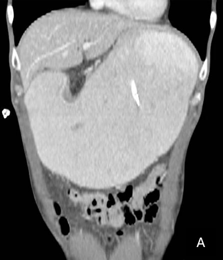
Figure: Coronal CT with diffuse proximal distension of the stomach, right upper quadrant malrotation, wall thickening of gastroduodenal junction.
Disclosures:
Hayes Walker indicated no relevant financial relationships.
Ronald Jordan indicated no relevant financial relationships.
Hayes Walker, MD, Ronald Scott. Jordan, MD. P5085 - Eosinophilic Gastritis Associated With Gastric Adenocarcinoma Presenting With Massive Gastric Outlet Obstruction, ACG 2024 Annual Scientific Meeting Abstracts. Philadelphia, PA: American College of Gastroenterology.
