Tuesday Poster Session
Category: Stomach
P5087 - A Case of Gastric Wall Abscess Presenting as Gastric Tumor
Tuesday, October 29, 2024
10:30 AM - 4:00 PM ET
Location: Exhibit Hall E

Has Audio

Hyder Alikhan, BA
Cooper Medical School of Rowan University
Camden, NJ
Presenting Author(s)
Hyder Alikhan, BA1, Kathy N. Williams, MD, MS2, Kunal Dalal, MD3
1Cooper Medical School of Rowan University, Camden, NJ; 2Cooper University Hospital, Philadelphia, PA; 3Cooper University Hospital, Camden, NJ
Introduction: Gastric wall abscesses (GWA) are rare pyogenic collections that occur within the gastric wall. They have been previously reported in older individuals and can mimic gastric cancers. We present a unique case of a patient with previous gastric ulcer perforation presenting with delayed onset of a gastric collection that was found to be a GWA and phlegmon formation after total gastrectomy.
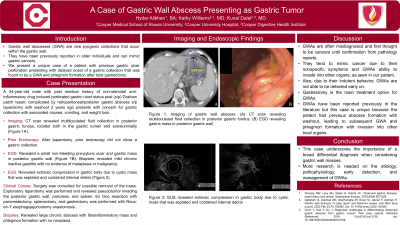
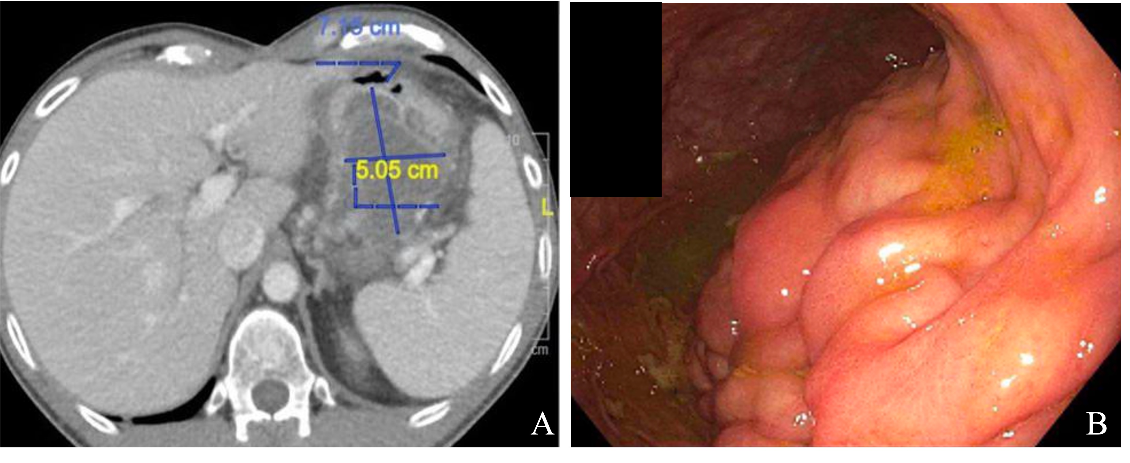
Case Description/Methods: A 34-year-old male with past medical history of non-steroidal anti-inflammatory drug induced perforated gastric ulcer status post (s/p) Graham patch repair, complicated by retroperitoneal/posterior gastric abscess s/p laparotomy with washout 2 years ago presents with concern for gastric collection with associated nausea, vomiting, and weight loss. CT scan revealed multiloculated fluid collection in posterior gastric fundus, located both in the gastric lumen and extraluminally (Figure 1A). Prior endoscopy after laparotomy did not show a gastric collection. EGD revealed a small non-bleeding pre-pyloric ulcer and gastric mass in posterior gastric wall (Figure 1B). Biopsies revealed mild chronic inactive gastritis with no evidence of metaplasia or malignancy. EUS revealed extrinsic compression in gastric body due to cystic mass that was septated and contained internal debris. Surgery was consulted for possible removal of the mass. Exploratory laparotomy was performed and revealed pseudotumor invading the posterior gastric wall, pancreas, and spleen. En bloc resection with pancreatectomy, splenectomy, and gastrectomy was performed with Roux-en-Y esophagojejunostomy anastomosis. Biopsies revealed large chronic abscess with fibroinflammatory mass and phlegmon formation with no neoplasia.
Discussion: GWAs are often misdiagnosed and first thought to be cancers until confirmation from pathology reports. Due to nonspecific symptoms and GWAs ability to invade into other organs, as seen in our patient, they tend to mimic cancer. However, due to their indolent behavior, GWAs are not able to be detected early on. Gastrectomy is the main treatment option for GWAs. GWAs have been reported previously in the literature; however, this case is unique because the patient had previous abscess formation with washout, with subsequent GWA and phlegmon formation with invasion into other local organs. This case underscores the importance of a broad differential diagnosis when considering gastric wall masses. More research is needed on the etiology, pathophysiology, early detection, and management of GWAs.

Disclosures:
Hyder Alikhan, BA1, Kathy N. Williams, MD, MS2, Kunal Dalal, MD3. P5087 - A Case of Gastric Wall Abscess Presenting as Gastric Tumor, ACG 2024 Annual Scientific Meeting Abstracts. Philadelphia, PA: American College of Gastroenterology.
1Cooper Medical School of Rowan University, Camden, NJ; 2Cooper University Hospital, Philadelphia, PA; 3Cooper University Hospital, Camden, NJ
Introduction: Gastric wall abscesses (GWA) are rare pyogenic collections that occur within the gastric wall. They have been previously reported in older individuals and can mimic gastric cancers. We present a unique case of a patient with previous gastric ulcer perforation presenting with delayed onset of a gastric collection that was found to be a GWA and phlegmon formation after total gastrectomy.
Case Description/Methods: A 34-year-old male with past medical history of non-steroidal anti-inflammatory drug induced perforated gastric ulcer status post (s/p) Graham patch repair, complicated by retroperitoneal/posterior gastric abscess s/p laparotomy with washout 2 years ago presents with concern for gastric collection with associated nausea, vomiting, and weight loss. CT scan revealed multiloculated fluid collection in posterior gastric fundus, located both in the gastric lumen and extraluminally (Figure 1A). Prior endoscopy after laparotomy did not show a gastric collection. EGD revealed a small non-bleeding pre-pyloric ulcer and gastric mass in posterior gastric wall (Figure 1B). Biopsies revealed mild chronic inactive gastritis with no evidence of metaplasia or malignancy. EUS revealed extrinsic compression in gastric body due to cystic mass that was septated and contained internal debris. Surgery was consulted for possible removal of the mass. Exploratory laparotomy was performed and revealed pseudotumor invading the posterior gastric wall, pancreas, and spleen. En bloc resection with pancreatectomy, splenectomy, and gastrectomy was performed with Roux-en-Y esophagojejunostomy anastomosis. Biopsies revealed large chronic abscess with fibroinflammatory mass and phlegmon formation with no neoplasia.
Discussion: GWAs are often misdiagnosed and first thought to be cancers until confirmation from pathology reports. Due to nonspecific symptoms and GWAs ability to invade into other organs, as seen in our patient, they tend to mimic cancer. However, due to their indolent behavior, GWAs are not able to be detected early on. Gastrectomy is the main treatment option for GWAs. GWAs have been reported previously in the literature; however, this case is unique because the patient had previous abscess formation with washout, with subsequent GWA and phlegmon formation with invasion into other local organs. This case underscores the importance of a broad differential diagnosis when considering gastric wall masses. More research is needed on the etiology, pathophysiology, early detection, and management of GWAs.

Figure: Figure 1. Imaging of gastric wall abscess. (A) CT scan revealing multiloculated fluid collection in posterior gastric fundus. (B) EGD revealing gastric mass in posterior gastric wall
Disclosures:
Hyder Alikhan indicated no relevant financial relationships.
Kathy Williams indicated no relevant financial relationships.
Kunal Dalal indicated no relevant financial relationships.
Hyder Alikhan, BA1, Kathy N. Williams, MD, MS2, Kunal Dalal, MD3. P5087 - A Case of Gastric Wall Abscess Presenting as Gastric Tumor, ACG 2024 Annual Scientific Meeting Abstracts. Philadelphia, PA: American College of Gastroenterology.
