Tuesday Poster Session
Category: Liver
P4820 - Non-Gestational Choriocarcinoma Presenting as a Liver Mass in a Postmenopausal Woman
Tuesday, October 29, 2024
10:30 AM - 4:00 PM ET
Location: Exhibit Hall E

Has Audio

Katherine O. Jones, DO
UCSF Fresno
Fresno, CA
Presenting Author(s)
Katherine O. Jones, DO, Arpine Petrosyan, MD, Jennifer Yoon, MD, Marina Roytman, MD
UCSF Fresno, Fresno, CA
Introduction: Choriocarcinoma is a rare, aggressive malignancy characterized by trophoblastic disease and defined by evidence of cytotrophoblastic and syncytiotrophoblastic cells on immunohistochemical analysis. Choriocarcinoma is divided into two main categories, gestational and non-gestational. It typically presents within one year of a preceding pregnancy, with the majority of documented cases being intrauterine and gestational in origin. As such, the condition is often associated with younger reproductive age women, with the average age of onset from 12 to 25 years. The incidence of non-gestational choriocarcinoma is difficult to assess as few cases have been reported in the current literature. Estimates suggest non-gestational choriocarcinoma accounts for 0.6% of all malignant ovarian germ cell tumors.
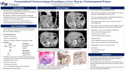
Case Description/Methods: An 82-year-old woman with history of chronic epigastric pain and hysterectomy presented to the emergency department with generalized weakness, abdominal pain, and 25-pound weight loss over a 6-month period. Workup was notable for normocytic anemia, without overt signs of active bleeding at the time of presentation. Computed tomography abdomen and pelvis with contrast revealed multiple liver masses, the two largest measuring 10x8.5x9cm and 7x6x5cm. Alpha fetoprotein and serum human chorionic gonadotropin levels were notably elevated to 82.5 ng/mL (reference: 0.0 - 8.0 ng/mL) and 170,739 m[IU]/mL (reference: 0.0 - 5.0 m[IU]/mL), respectively. Follow up ultrasound guided biopsy of the liver and immunohistochemical analysis revealed a high-grade malignant neoplasm positive for cytokeratin, GATA-3, p63 (cytotrophoblast cells), glypican-3, and inhibin (syncytiotrophoblast cells). Overall, the morphologic appearance, immunophenotype, and elevated tumor markers were consistent with the diagnosis of choriocarcinoma. Given her age and advanced disease the patient decided to defer further workup and chose to pursue hospice.
Discussion: Non-gestational choriocarcinoma presents unique diagnostic challenges, especially in the post-menopausal population where trophoblastic disease is typically excluded from the differential. Here we highlight key features of this aggressive malignancy in conjunction with the patient’s unique clinical presentation as an 82-year-old postmenopausal female with history of prior hysterectomy. Our case also emphasizes the importance of having this rare diagnosis in mind when finding metastatic lesions within the liver.

Disclosures:
Katherine O. Jones, DO, Arpine Petrosyan, MD, Jennifer Yoon, MD, Marina Roytman, MD. P4820 - Non-Gestational Choriocarcinoma Presenting as a Liver Mass in a Postmenopausal Woman, ACG 2024 Annual Scientific Meeting Abstracts. Philadelphia, PA: American College of Gastroenterology.
UCSF Fresno, Fresno, CA
Introduction: Choriocarcinoma is a rare, aggressive malignancy characterized by trophoblastic disease and defined by evidence of cytotrophoblastic and syncytiotrophoblastic cells on immunohistochemical analysis. Choriocarcinoma is divided into two main categories, gestational and non-gestational. It typically presents within one year of a preceding pregnancy, with the majority of documented cases being intrauterine and gestational in origin. As such, the condition is often associated with younger reproductive age women, with the average age of onset from 12 to 25 years. The incidence of non-gestational choriocarcinoma is difficult to assess as few cases have been reported in the current literature. Estimates suggest non-gestational choriocarcinoma accounts for 0.6% of all malignant ovarian germ cell tumors.
Case Description/Methods: An 82-year-old woman with history of chronic epigastric pain and hysterectomy presented to the emergency department with generalized weakness, abdominal pain, and 25-pound weight loss over a 6-month period. Workup was notable for normocytic anemia, without overt signs of active bleeding at the time of presentation. Computed tomography abdomen and pelvis with contrast revealed multiple liver masses, the two largest measuring 10x8.5x9cm and 7x6x5cm. Alpha fetoprotein and serum human chorionic gonadotropin levels were notably elevated to 82.5 ng/mL (reference: 0.0 - 8.0 ng/mL) and 170,739 m[IU]/mL (reference: 0.0 - 5.0 m[IU]/mL), respectively. Follow up ultrasound guided biopsy of the liver and immunohistochemical analysis revealed a high-grade malignant neoplasm positive for cytokeratin, GATA-3, p63 (cytotrophoblast cells), glypican-3, and inhibin (syncytiotrophoblast cells). Overall, the morphologic appearance, immunophenotype, and elevated tumor markers were consistent with the diagnosis of choriocarcinoma. Given her age and advanced disease the patient decided to defer further workup and chose to pursue hospice.
Discussion: Non-gestational choriocarcinoma presents unique diagnostic challenges, especially in the post-menopausal population where trophoblastic disease is typically excluded from the differential. Here we highlight key features of this aggressive malignancy in conjunction with the patient’s unique clinical presentation as an 82-year-old postmenopausal female with history of prior hysterectomy. Our case also emphasizes the importance of having this rare diagnosis in mind when finding metastatic lesions within the liver.

Figure: A) Multiple hyper-vascular and irregular masses
B) Lesional process, with evidence of background hemorrhage
C) IHC positive for inhibin (syncytiotrophoblast cells)
B) Lesional process, with evidence of background hemorrhage
C) IHC positive for inhibin (syncytiotrophoblast cells)
Disclosures:
Katherine Jones indicated no relevant financial relationships.
Arpine Petrosyan indicated no relevant financial relationships.
Jennifer Yoon indicated no relevant financial relationships.
Marina Roytman: Gilead – Speakers Bureau. Madrigal – Speakers Bureau. Salix – Speakers Bureau.
Katherine O. Jones, DO, Arpine Petrosyan, MD, Jennifer Yoon, MD, Marina Roytman, MD. P4820 - Non-Gestational Choriocarcinoma Presenting as a Liver Mass in a Postmenopausal Woman, ACG 2024 Annual Scientific Meeting Abstracts. Philadelphia, PA: American College of Gastroenterology.

