Tuesday Poster Session
Category: Liver
P4821 - A Case of a Hearty Liver Cyst
Tuesday, October 29, 2024
10:30 AM - 4:00 PM ET
Location: Exhibit Hall E

Has Audio
- MS
Michelle Shi, MD
North Shore University Hospital - Northwell Health
Manhasset, New York
Presenting Author(s)
Ethan Berman, MD1, Michelle Shi, MD2, Hatef Massoumi, MD2
1New York University Langone Health, New York, NY; 2North Shore University Hospital - Northwell Health, Manhasset, NY
Introduction: Hepatic cysts are common and are generally found incidentally on imaging studies performed for unrelated reasons. They are usually solitary, asymptomatic, and do not require interventions. Rarely, a cyst can become large enough to cause symptoms such as pain or affecting adjacent organs. In this unusual case, our patient presented with bi-ventricular heart failure secondary to extrinsic compression from a giant hepatic cyst.
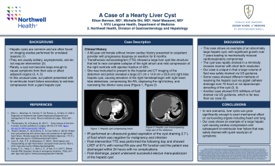
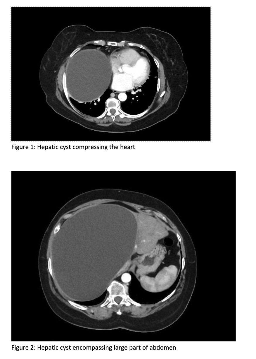
Case Description/Methods: A 66-year-old female without known cardiac history presented to outpatient provider with progressive dyspnea on exertion for 8 months and early satiety for 6 months. Outpatient transthoracic echocardiogram (TTE) showed a large liver cyst-like structure that led to near complete collapse of the right atrium (RA) and mild compression of the right ventricle (RV). The patient was known to have an asymptomatic hepatic cyst since about 12 years ago. She subsequently presented to outside hospital for further evaluation, and CT imaging revealed a large (21 cm x 14.9 cm x 22.8 cm) right lobe hepatic cyst, compressing the RA, displacing the right kidney, and narrowing the inferior vena cava (Figure 1, Figure 2). The patient was then transferred to our center for escalation of care. Inpatient TTE was notable for left ventricular systolic ejection fraction (LVEF) of 49%, extrinsic compression of RA and reduced RV systolic function. The interventional radiology team performed a US-guided aspiration of the hepatic cyst, draining 2.7 liters of slightly cloudy clear fluid with significant decrease in the size of the hepatic cyst. The cyst fluid was negative for signs of infection or malignancy. Post-intervention TTE showed LVEF of 61% with normal RA size and RV function. The patient was discharged within 24 hours of the hepatic cyst aspiration with stable hemodynamics. One-month post-discharge, patient underwent successful elective marsupialization of the hepatic cyst, with additional 2 liters of serous fluid aspirated.
Discussion: This case shows an example of an abnormally large symptomatic hepatic cyst rapidly drained in a minimally invasive manner with short-term resolution. The patient had no complications from the procedure and was discharged within 24 hours of drainage. She had complete resolution of the symptoms and remained asymptomatic until the eventual surgical intervention. Our case is unique in that a large volume of fluid was safely drained via US guidance.

Disclosures:
Ethan Berman, MD1, Michelle Shi, MD2, Hatef Massoumi, MD2. P4821 - A Case of a Hearty Liver Cyst, ACG 2024 Annual Scientific Meeting Abstracts. Philadelphia, PA: American College of Gastroenterology.
1New York University Langone Health, New York, NY; 2North Shore University Hospital - Northwell Health, Manhasset, NY
Introduction: Hepatic cysts are common and are generally found incidentally on imaging studies performed for unrelated reasons. They are usually solitary, asymptomatic, and do not require interventions. Rarely, a cyst can become large enough to cause symptoms such as pain or affecting adjacent organs. In this unusual case, our patient presented with bi-ventricular heart failure secondary to extrinsic compression from a giant hepatic cyst.
Case Description/Methods: A 66-year-old female without known cardiac history presented to outpatient provider with progressive dyspnea on exertion for 8 months and early satiety for 6 months. Outpatient transthoracic echocardiogram (TTE) showed a large liver cyst-like structure that led to near complete collapse of the right atrium (RA) and mild compression of the right ventricle (RV). The patient was known to have an asymptomatic hepatic cyst since about 12 years ago. She subsequently presented to outside hospital for further evaluation, and CT imaging revealed a large (21 cm x 14.9 cm x 22.8 cm) right lobe hepatic cyst, compressing the RA, displacing the right kidney, and narrowing the inferior vena cava (Figure 1, Figure 2). The patient was then transferred to our center for escalation of care. Inpatient TTE was notable for left ventricular systolic ejection fraction (LVEF) of 49%, extrinsic compression of RA and reduced RV systolic function. The interventional radiology team performed a US-guided aspiration of the hepatic cyst, draining 2.7 liters of slightly cloudy clear fluid with significant decrease in the size of the hepatic cyst. The cyst fluid was negative for signs of infection or malignancy. Post-intervention TTE showed LVEF of 61% with normal RA size and RV function. The patient was discharged within 24 hours of the hepatic cyst aspiration with stable hemodynamics. One-month post-discharge, patient underwent successful elective marsupialization of the hepatic cyst, with additional 2 liters of serous fluid aspirated.
Discussion: This case shows an example of an abnormally large symptomatic hepatic cyst rapidly drained in a minimally invasive manner with short-term resolution. The patient had no complications from the procedure and was discharged within 24 hours of drainage. She had complete resolution of the symptoms and remained asymptomatic until the eventual surgical intervention. Our case is unique in that a large volume of fluid was safely drained via US guidance.

Figure: CT Images of the hepatic cyst
Disclosures:
Ethan Berman indicated no relevant financial relationships.
Michelle Shi indicated no relevant financial relationships.
Hatef Massoumi indicated no relevant financial relationships.
Ethan Berman, MD1, Michelle Shi, MD2, Hatef Massoumi, MD2. P4821 - A Case of a Hearty Liver Cyst, ACG 2024 Annual Scientific Meeting Abstracts. Philadelphia, PA: American College of Gastroenterology.
