Tuesday Poster Session
Category: Liver
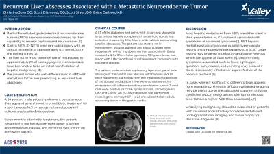
P4757 - Recurrent Liver Abscesses Associated With a Metastatic Neuroendocrine Tumor
Tuesday, October 29, 2024
10:30 AM - 4:00 PM ET
Location: Exhibit Hall E

Has Audio

Christina Joya, DO
Valley Hospital Medical Center
Las Vegas, NV
Presenting Author(s)
Christina Joya, DO, Scott Diamond, DO, Brian Carlson, MD
Valley Hospital Medical Center, Las Vegas, NV
Introduction: Well-differentiated gastrointestinal neuroendocrine tumors (NETs) are neoplasms characterized by their capability to secrete peptides and neuroamines. Gastric NETs (G-NETs) are a rare subcategory with an annual incidence of approximately 0.17 per 10,000 in the United States. The liver is the most common site of metastases. In approximately 2% of cases, pyogenic liver abscesses have been noted to be an initial manifestation of hepatic malignancy. We present a case of a well-differentiated G-NET with metastasis to the liver presenting as recurrent liver abscesses.
Case Description/Methods: A 54 year old male underwent percutaneous drainage and several months of antibiotic treatment for a spontaneous 14.9 cm pyogenic liver abscess with cultures positive for Enterobacter. Seven months after initial treatment, the patient presented with RUQ abdominal pain, nausea, and vomiting. WBC on admission was 9.13. A CT of the abdomen and pelvis with IV contrast showed a large central hepatic complex with an air-fluid containing collection measuring 9.6 x 8.4 cm and multiple surrounding satellite abscesses. Wound, aspirate, and blood cultures were negative. An MRI of the abdomen liver protocol with Eovist revealed a 12.1 x 11.1 cm heterogeneous T2 hyperintense liver lesion with a thickened wall of enhancement.
The patient underwent wide drainage of the central liver abscess with biopsies and JP drain placement. Pathology from the biopsies of the abscess and adjacent liver were consistent with a metastatic well-differentiated NET. Tumor cells were positive for CD56, synaptophysin, chromogranin, CK7, and CAM5. An EGD with biopsies was performed revealing the primary NET – a 2.5 cm subepithelial nodular appearing lesion in the gastric cardia.
Discussion: Most hepatic metastases from NETs are either silent in their presentation or, if functional, associated with symptoms of carcinoid syndrome. NET hepatic metastases appear as solid-hypervascular lesions on CT. Large lesions may undergo liquefaction and tumor necrosis which can appear as fluid levels. Symptoms associated such as fever, RUQ pain, nausea, and vomiting may present if there is secondary infection or superinfection of the necrotic material. In cases where it is difficult to differentiate an abscess from malignancy, MRI with diffusion-weighted-imaging may be useful. Underlying malignancy should be suspected in patients with recurrent and refractory abscesses. Additional imaging and tissue biopsy is warranted for definitive diagnosis.
Disclosures:
Christina Joya, DO, Scott Diamond, DO, Brian Carlson, MD. P4757 - Recurrent Liver Abscesses Associated With a Metastatic Neuroendocrine Tumor, ACG 2024 Annual Scientific Meeting Abstracts. Philadelphia, PA: American College of Gastroenterology.
Valley Hospital Medical Center, Las Vegas, NV
Introduction: Well-differentiated gastrointestinal neuroendocrine tumors (NETs) are neoplasms characterized by their capability to secrete peptides and neuroamines. Gastric NETs (G-NETs) are a rare subcategory with an annual incidence of approximately 0.17 per 10,000 in the United States. The liver is the most common site of metastases. In approximately 2% of cases, pyogenic liver abscesses have been noted to be an initial manifestation of hepatic malignancy. We present a case of a well-differentiated G-NET with metastasis to the liver presenting as recurrent liver abscesses.
Case Description/Methods: A 54 year old male underwent percutaneous drainage and several months of antibiotic treatment for a spontaneous 14.9 cm pyogenic liver abscess with cultures positive for Enterobacter. Seven months after initial treatment, the patient presented with RUQ abdominal pain, nausea, and vomiting. WBC on admission was 9.13. A CT of the abdomen and pelvis with IV contrast showed a large central hepatic complex with an air-fluid containing collection measuring 9.6 x 8.4 cm and multiple surrounding satellite abscesses. Wound, aspirate, and blood cultures were negative. An MRI of the abdomen liver protocol with Eovist revealed a 12.1 x 11.1 cm heterogeneous T2 hyperintense liver lesion with a thickened wall of enhancement.
The patient underwent wide drainage of the central liver abscess with biopsies and JP drain placement. Pathology from the biopsies of the abscess and adjacent liver were consistent with a metastatic well-differentiated NET. Tumor cells were positive for CD56, synaptophysin, chromogranin, CK7, and CAM5. An EGD with biopsies was performed revealing the primary NET – a 2.5 cm subepithelial nodular appearing lesion in the gastric cardia.
Discussion: Most hepatic metastases from NETs are either silent in their presentation or, if functional, associated with symptoms of carcinoid syndrome. NET hepatic metastases appear as solid-hypervascular lesions on CT. Large lesions may undergo liquefaction and tumor necrosis which can appear as fluid levels. Symptoms associated such as fever, RUQ pain, nausea, and vomiting may present if there is secondary infection or superinfection of the necrotic material. In cases where it is difficult to differentiate an abscess from malignancy, MRI with diffusion-weighted-imaging may be useful. Underlying malignancy should be suspected in patients with recurrent and refractory abscesses. Additional imaging and tissue biopsy is warranted for definitive diagnosis.
Disclosures:
Christina Joya indicated no relevant financial relationships.
Scott Diamond indicated no relevant financial relationships.
Brian Carlson indicated no relevant financial relationships.
Christina Joya, DO, Scott Diamond, DO, Brian Carlson, MD. P4757 - Recurrent Liver Abscesses Associated With a Metastatic Neuroendocrine Tumor, ACG 2024 Annual Scientific Meeting Abstracts. Philadelphia, PA: American College of Gastroenterology.
