Tuesday Poster Session
Category: Liver
P4758 - Weight Loss is the Boss: Hepatic Adenomas
Tuesday, October 29, 2024
10:30 AM - 4:00 PM ET
Location: Exhibit Hall E

Has Audio

Christopher J. Cieker, MD
University of Texas Health San Antonio
San Antonio, TX
Presenting Author(s)
Christopher J. Cieker, MD1, Eugenia Tsai, MD2, Lisa Pedicone, PhD2, Andres Gomez-Aldana, MD2, Jan Petrasek, MD, PhD2, Carmen Landaverde, MD3, Eric J. Lawitz, MD4, Fred Poordad, MD2, Fabian Rodas, MD2
1University of Texas Health San Antonio, San Antonio, TX; 2Texas Liver Institute, San Antonio, TX; 3Texas Liver Institute, Austin, TX; 4The Texas Liver Institute, University of Texas Health, San Antonio, TX
Introduction: A hepatocellular adenoma (HCA) is a benign liver tumor with incidence of 1 case per million people.1 HCAs have a prevalence of 10-30 per million women on oral contraceptives (OCPs).1 We present a case where HCAs were managed indirectly by weight loss with semaglutide, a glucagon-like peptide-1 (GLP-1A) receptor agonist.
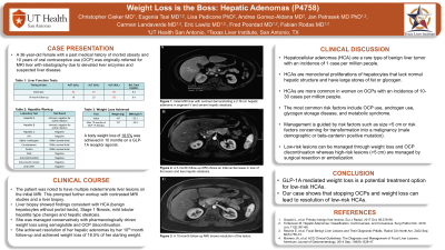
Case Description/Methods: A 36-year-old obese female (body mass index of 53) with 10 years of OCP use was seen due to elevated liver enzymes from hepatic steatosis (HS) versus metabolic dysfunction-associated steatohepatitis (MASH). She started semaglutide for obesity and was referred for MRI. Findings showed moderate to severe HS (fat fraction of 20.7-22.8%) and at least 7 T2 bright liver masses reaching up to 3.4 cm (figure 1A shows a 2.76 cm lesion). Her OCP was stopped.
A liver biopsy showed findings consistent with HCA (benign hepatocytes without portal tracts), stage 1 fibrosis, mild lobular hepatitis-type changes, and background HS. This was consistent with a diagnosis of MASH with HCAs. Conservative treatment was chosen through imaging surveillance. Improvement was seen on 3-month follow-up MRI (figure 1B). Over 10 months, the patient lost 18.5% of her body weight (60 lbs) and had resolution of the HCAs and HS on MRI (figure 1C).
Discussion: HCAs are monoclonal proliferations of hepatocytes that lack normal hepatic structure and have large stores of fat or glycogen.1,2,3 HS is also due to fat accumulation from dysregulation of lipid metabolism. HCAs are affected by OCPs, metabolic syndrome, and androgen use.1,2,3 HCAs can cause life-threatening bleeds from ruptured lesions or turn into hepatocellular carcinoma.1,2,3 High risk lesions >5 cm are managed by surgical resection or embolization.4 Low risk lesions can be managed by stopping OCPs and weight loss.4
GLP-1A mediated weight loss is a potential treatment option for HCAs. Our case shows that stopping OCPs and weight loss can lead to resolution of HCAs.
References:
1. Grazioli L, et al. Primary benign liver lesions. Eur J Radiol. 2017 Oct; 95:378-98.
2. Torbenson M. Hepatic Adenomas: Classification, Controversies, and Consensus. Surg Pathol Clin. 2018 Jun; 11(2):351-66.
3. Reizine E, et al. Focal Benign Liver Lesions and Their Diagnostic Pitfalls. Radiol Clin North Am. 2022 Sep; 60(5):755-73.
4. Marrero JA, et al. ACG Clinical Guidelines: The Diagnosis and Management of Focal Liver Lesions. American Journal of Gastroenterology. 2014 Sep; 109(9):1328-47.

Disclosures:
Christopher J. Cieker, MD1, Eugenia Tsai, MD2, Lisa Pedicone, PhD2, Andres Gomez-Aldana, MD2, Jan Petrasek, MD, PhD2, Carmen Landaverde, MD3, Eric J. Lawitz, MD4, Fred Poordad, MD2, Fabian Rodas, MD2. P4758 - Weight Loss is the Boss: Hepatic Adenomas, ACG 2024 Annual Scientific Meeting Abstracts. Philadelphia, PA: American College of Gastroenterology.
1University of Texas Health San Antonio, San Antonio, TX; 2Texas Liver Institute, San Antonio, TX; 3Texas Liver Institute, Austin, TX; 4The Texas Liver Institute, University of Texas Health, San Antonio, TX
Introduction: A hepatocellular adenoma (HCA) is a benign liver tumor with incidence of 1 case per million people.1 HCAs have a prevalence of 10-30 per million women on oral contraceptives (OCPs).1 We present a case where HCAs were managed indirectly by weight loss with semaglutide, a glucagon-like peptide-1 (GLP-1A) receptor agonist.
Case Description/Methods: A 36-year-old obese female (body mass index of 53) with 10 years of OCP use was seen due to elevated liver enzymes from hepatic steatosis (HS) versus metabolic dysfunction-associated steatohepatitis (MASH). She started semaglutide for obesity and was referred for MRI. Findings showed moderate to severe HS (fat fraction of 20.7-22.8%) and at least 7 T2 bright liver masses reaching up to 3.4 cm (figure 1A shows a 2.76 cm lesion). Her OCP was stopped.
A liver biopsy showed findings consistent with HCA (benign hepatocytes without portal tracts), stage 1 fibrosis, mild lobular hepatitis-type changes, and background HS. This was consistent with a diagnosis of MASH with HCAs. Conservative treatment was chosen through imaging surveillance. Improvement was seen on 3-month follow-up MRI (figure 1B). Over 10 months, the patient lost 18.5% of her body weight (60 lbs) and had resolution of the HCAs and HS on MRI (figure 1C).
Discussion: HCAs are monoclonal proliferations of hepatocytes that lack normal hepatic structure and have large stores of fat or glycogen.1,2,3 HS is also due to fat accumulation from dysregulation of lipid metabolism. HCAs are affected by OCPs, metabolic syndrome, and androgen use.1,2,3 HCAs can cause life-threatening bleeds from ruptured lesions or turn into hepatocellular carcinoma.1,2,3 High risk lesions >5 cm are managed by surgical resection or embolization.4 Low risk lesions can be managed by stopping OCPs and weight loss.4
GLP-1A mediated weight loss is a potential treatment option for HCAs. Our case shows that stopping OCPs and weight loss can lead to resolution of HCAs.
References:
1. Grazioli L, et al. Primary benign liver lesions. Eur J Radiol. 2017 Oct; 95:378-98.
2. Torbenson M. Hepatic Adenomas: Classification, Controversies, and Consensus. Surg Pathol Clin. 2018 Jun; 11(2):351-66.
3. Reizine E, et al. Focal Benign Liver Lesions and Their Diagnostic Pitfalls. Radiol Clin North Am. 2022 Sep; 60(5):755-73.
4. Marrero JA, et al. ACG Clinical Guidelines: The Diagnosis and Management of Focal Liver Lesions. American Journal of Gastroenterology. 2014 Sep; 109(9):1328-47.

Figure: Figure 1. The initial MRI liver with contrast demonstrated (A) a 2.76 cm hepatic adenoma in Segment V and severe hepatic steatosis. A follow-up MRI liver with contrast approximately 3 months later displays (B) interval decrease in size of the lesion and improvement of hepatic steatosis. (C) A 10 month follow up contrasted study shows complete resolution of liver lesion.
Disclosures:
Christopher Cieker indicated no relevant financial relationships.
Eugenia Tsai indicated no relevant financial relationships.
Lisa Pedicone indicated no relevant financial relationships.
Andres Gomez-Aldana indicated no relevant financial relationships.
Jan Petrasek indicated no relevant financial relationships.
Carmen Landaverde indicated no relevant financial relationships.
Eric Lawitz: 89Bio – Consultant, Grant/Research Support. Akero – Consultant, Grant/Research Support. Alnylam – Consultant, Grant/Research Support. Amgen – Consultant, Grant/Research Support. AstraZeneca – Consultant, Grant/Research Support. Axcella Health – Consultant, Grant/Research Support. Boehringer Ingelheim – Consultant, Grant/Research Support. Bristol Myers Squibb – Consultant, Grant/Research Support. CymaBay, a Gilead Sciences Company – Consultant, Grant/Research Support. CytoDyn – Consultant, Grant/Research Support. DSM – Consultant, Grant/Research Support. Durect – Consultant, Grant/Research Support. Eli Lilly – Consultant, Grant/Research Support. Enanta – Consultant, Grant/Research Support. Enyo – Consultant, Grant/Research Support. Exalenz – Consultant, Grant/Research Support. Galectin – Consultant, Grant/Research Support. Galmed – Consultant, Grant/Research Support. Genentech – Consultant, Grant/Research Support. Genfit – Consultant, Grant/Research Support. Gilead Sciences, Inc. – Consultant, Grant/Research Support. GSK – Consultant, Grant/Research Support. Hanmi – Consultant, Grant/Research Support. Hightide – Consultant, Grant/Research Support. Intercept – Consultant, Grant/Research Support. Inventiva – Consultant, Grant/Research Support. Ipsen – Consultant, Grant/Research Support. Janssen – Consultant, Grant/Research Support. Madrigal – Consultant, Grant/Research Support. Merck & Co – Consultant, Grant/Research Support. NGM – Consultant, Grant/Research Support. Northsea – Consultant, Grant/Research Support. Novartis – Consultant, Grant/Research Support. Novo Nordisk – Consultant, Grant/Research Support. Pfizer – Consultant, Grant/Research Support. Poxel Co – Consultant, Grant/Research Support. Roche – Consultant, Grant/Research Support. Sagimet – Consultant, Grant/Research Support. Terns – Consultant, Grant/Research Support. Viking – Consultant, Grant/Research Support. Zydus – Consultant, Grant/Research Support.
Fred Poordad indicated no relevant financial relationships.
Fabian Rodas indicated no relevant financial relationships.
Christopher J. Cieker, MD1, Eugenia Tsai, MD2, Lisa Pedicone, PhD2, Andres Gomez-Aldana, MD2, Jan Petrasek, MD, PhD2, Carmen Landaverde, MD3, Eric J. Lawitz, MD4, Fred Poordad, MD2, Fabian Rodas, MD2. P4758 - Weight Loss is the Boss: Hepatic Adenomas, ACG 2024 Annual Scientific Meeting Abstracts. Philadelphia, PA: American College of Gastroenterology.

