Tuesday Poster Session
Category: Liver
P4764 - A Peculiar Case of Hepatolithiasis in the Western World
Tuesday, October 29, 2024
10:30 AM - 4:00 PM ET
Location: Exhibit Hall E

Has Audio

Shivani Sreenivasan, MBBS
Long Island Jewish Medical Center, Forest Hills- Northwell Health
Forest Hills, NY
Presenting Author(s)
Shivani Sreenivasan, MBBS1, WonKyung Cho, MD, MPH2, Heng K. Nhoung, MD3, Pan Derek, DO4, Henry Jen, MD5, Timea Csak, MD4
1Long Island Jewish Medical Center, Forest Hills- Northwell Health, Forest Hills, NY; 2Long Island Jewish Medical Center, Forest Hills - Northwell Health, New York, NY; 3Northwell Health, Long Island City, NY; 4Long Island Jewish Medical Center, Forest Hills- Northwell Health, New York, NY; 5Long Island Jewish Medical Center, Forest Hills - Northwell Health, Forest Hills, NY
Introduction: Hepatolithiasis is the presence of biliary stones in the intrahepatic bile ducts above the hilar confluence. It presents as recurrent upper abdominal pain and can be complicated by recurrent pyogenic cholangitis. It is a risk factor for intrahepatic cholangiocarcinoma. Treatment aims to prevent cholangitis and halt disease progression to hepato-biliary fibrosis. Although common in Southeast Asia, it is rare in the West with a prevalence of 1% and rarely presents before the 5th decade. Stone burden predominantly affects the left hepatic lobe. This is a rare presentation of hepatolithiasis in a young female in the United States.
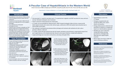
Case Description/Methods: Patient is a 26-year-old Hispanic female who presented with recurrent epigastric pain. Magnetic resonance cholangiopancreatography (MRCP) showed extensive intra and extrahepatic calculi confirmed on endoscopic ultrasound. Endoscopic retrograde cholangiopancreatography (ERCP) was performed for partial stone clearance and stent placement. ERCP was repeated in 2 weeks for abdominal pain and CT abdomen suspicious for cholangitis. Small intrahepatic calculi remained despite balloon extraction and stent exchange. She presented in 1 month for persistent pain. CT suspected stent migration and ERCP was done for stone and stent removal. Multiple intrahepatic calculi remained. Patient underwent cholecystectomy for cholecystitis. In 2 weeks, she was admitted for abdominal pain. MRCP showed intrahepatic biliary stricture, numerous left lateral segment stones and a stone at the stricture apex, confirmed on ERCP with partial stone extraction due to stone burden and location. She underwent transhepatic percutaneous biliary drainage and treatment with antibiotics for bile culture growing C.albicans, E.faecium. In 3 weeks, partial left lobe hepatectomy with left hepatic ductotomy was performed, complicated by sepsis from post-surgical abscess at the hepatectomy bed. Abscess culture grew E.faecium, treated with antibiotics. Surgical pathology was inconsistent with primary sclerosing cholangitis (PSC).
Discussion: Hepatolithiasis is rare in the 2nd decade. Etiologies are genetic bile acid transporter defect, PSC, Caroli’s disease and parasites. Etiology determination, treatment and prevention of infection is complex and requires an interdisciplinary approach. Modalities for stone eradication include ERCP-biliary stenting, percutaneous cholangioscopic lithotripsy and surgical hepatic resection. Rate of stone recurrence is high and requires lifelong surveillance.
Disclosures:
Shivani Sreenivasan, MBBS1, WonKyung Cho, MD, MPH2, Heng K. Nhoung, MD3, Pan Derek, DO4, Henry Jen, MD5, Timea Csak, MD4. P4764 - A Peculiar Case of Hepatolithiasis in the Western World, ACG 2024 Annual Scientific Meeting Abstracts. Philadelphia, PA: American College of Gastroenterology.
1Long Island Jewish Medical Center, Forest Hills- Northwell Health, Forest Hills, NY; 2Long Island Jewish Medical Center, Forest Hills - Northwell Health, New York, NY; 3Northwell Health, Long Island City, NY; 4Long Island Jewish Medical Center, Forest Hills- Northwell Health, New York, NY; 5Long Island Jewish Medical Center, Forest Hills - Northwell Health, Forest Hills, NY
Introduction: Hepatolithiasis is the presence of biliary stones in the intrahepatic bile ducts above the hilar confluence. It presents as recurrent upper abdominal pain and can be complicated by recurrent pyogenic cholangitis. It is a risk factor for intrahepatic cholangiocarcinoma. Treatment aims to prevent cholangitis and halt disease progression to hepato-biliary fibrosis. Although common in Southeast Asia, it is rare in the West with a prevalence of 1% and rarely presents before the 5th decade. Stone burden predominantly affects the left hepatic lobe. This is a rare presentation of hepatolithiasis in a young female in the United States.
Case Description/Methods: Patient is a 26-year-old Hispanic female who presented with recurrent epigastric pain. Magnetic resonance cholangiopancreatography (MRCP) showed extensive intra and extrahepatic calculi confirmed on endoscopic ultrasound. Endoscopic retrograde cholangiopancreatography (ERCP) was performed for partial stone clearance and stent placement. ERCP was repeated in 2 weeks for abdominal pain and CT abdomen suspicious for cholangitis. Small intrahepatic calculi remained despite balloon extraction and stent exchange. She presented in 1 month for persistent pain. CT suspected stent migration and ERCP was done for stone and stent removal. Multiple intrahepatic calculi remained. Patient underwent cholecystectomy for cholecystitis. In 2 weeks, she was admitted for abdominal pain. MRCP showed intrahepatic biliary stricture, numerous left lateral segment stones and a stone at the stricture apex, confirmed on ERCP with partial stone extraction due to stone burden and location. She underwent transhepatic percutaneous biliary drainage and treatment with antibiotics for bile culture growing C.albicans, E.faecium. In 3 weeks, partial left lobe hepatectomy with left hepatic ductotomy was performed, complicated by sepsis from post-surgical abscess at the hepatectomy bed. Abscess culture grew E.faecium, treated with antibiotics. Surgical pathology was inconsistent with primary sclerosing cholangitis (PSC).
Discussion: Hepatolithiasis is rare in the 2nd decade. Etiologies are genetic bile acid transporter defect, PSC, Caroli’s disease and parasites. Etiology determination, treatment and prevention of infection is complex and requires an interdisciplinary approach. Modalities for stone eradication include ERCP-biliary stenting, percutaneous cholangioscopic lithotripsy and surgical hepatic resection. Rate of stone recurrence is high and requires lifelong surveillance.
Disclosures:
Shivani Sreenivasan indicated no relevant financial relationships.
WonKyung Cho indicated no relevant financial relationships.
Heng Nhoung indicated no relevant financial relationships.
Pan Derek indicated no relevant financial relationships.
Henry Jen indicated no relevant financial relationships.
Timea Csak indicated no relevant financial relationships.
Shivani Sreenivasan, MBBS1, WonKyung Cho, MD, MPH2, Heng K. Nhoung, MD3, Pan Derek, DO4, Henry Jen, MD5, Timea Csak, MD4. P4764 - A Peculiar Case of Hepatolithiasis in the Western World, ACG 2024 Annual Scientific Meeting Abstracts. Philadelphia, PA: American College of Gastroenterology.
