Tuesday Poster Session
Category: Interventional Endoscopy
P4520 - A Case Series of Threes: Three Gastrointestinal Carcinoid Tumors in Three Locations Treated With Three Novel Endoscopic Techniques
Tuesday, October 29, 2024
10:30 AM - 4:00 PM ET
Location: Exhibit Hall E

Has Audio

Amanda Jacubowsky, DO
Lehigh Valley Health Network
Allentown, PA
Presenting Author(s)
Amanda Jacubowsky, DO1, Chandler Patton, DO1, Timothy Graziano, DO1, Shashin Shah, MD2
1Lehigh Valley Health Network, Allentown, PA; 2Eastern Pennsylvania Gastrointestinal and Liver Specialists, Allentown, PA
Introduction: Carcinoid is a rare, slow-growing neuroendocrine tumor with an annual incidence of 5.25 per 100,000 patients. The treatment for non-metastatic disease has historically been surgical resection. Here, we present a case series of three patients with incidentally discovered carcinoid tumors that were treated with three novel, minimally invasive endoscopic procedures.
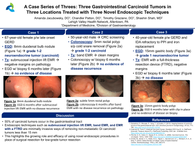
Case Description/Methods: First, a 67-year-old female presented with late-onset gastroesophageal reflux disease (GERD). An esophagogastroduodenoscopy (EGD) revealed an 8mm duodenal bulb nodule that was biopsied. Pathology showed a grade 1-2 neuroendocrine tumor. She subsequently under submucosal injection lift endoscopic mucosal resection (EMR) of the duodenal carcinoid with negative margins. A surveillance EGD 6 months later showed no evidence of disease recurrence with negative biopsies taken from the duodenal scar. Second, a 50-year-old male presented for a screening colonoscopy, which yielded a 5mm rectal polyp that was removed with a cold snare. Pathology revealed a well-differentiated, grade 1-2 carcinoid tumor requiring additional band EMR with clean margins. Repeat surveillance colonoscopy with biopsy demonstrated no residual rectal carcinoid. Third, a 40-year-old female presented for diagnostic EGD in the setting of GERD and iron deficiency anemia refractory to proton pump inhibitor therapy and iron supplementation. An EGD showed a 15mm nodule in the gastric body and pathology was consistent with a grade 1 neuroendocrine tumor. She underwent EMR with a full-thickness resection device (FTRD) used to remove the 15mm lesion. Pathology revealed negative margins for tumor invasion. A surveillance EGD 6 months later was negative for disease recurrence.
Discussion: About 55% of carcinoid tumors occur in the GI tract, most commonly in the rectum (34%), small bowel (26%), and stomach (12%). About 90% of patients are asymptomatic until the detection of advanced and metastatic changes, posing a significant diagnostic challenge. Endoscopic techniques such as submucosal injection lift EMR, band EMR, and EMR with a FTRD are minimally invasive ways of removing non-metastatic GI carcinoid tumors less than 15 mm. In this case series, all three patients had negative margins on the pathology report from the initial endoscopic removal of the tumors. Further, surveillance endoscopies showed no tumor recurrence. This highlights the growing role and efficacy of using novel endoscopic procedures in place of surgery for low-grade tumor resection.
Disclosures:
Amanda Jacubowsky, DO1, Chandler Patton, DO1, Timothy Graziano, DO1, Shashin Shah, MD2. P4520 - A Case Series of Threes: Three Gastrointestinal Carcinoid Tumors in Three Locations Treated With Three Novel Endoscopic Techniques, ACG 2024 Annual Scientific Meeting Abstracts. Philadelphia, PA: American College of Gastroenterology.
1Lehigh Valley Health Network, Allentown, PA; 2Eastern Pennsylvania Gastrointestinal and Liver Specialists, Allentown, PA
Introduction: Carcinoid is a rare, slow-growing neuroendocrine tumor with an annual incidence of 5.25 per 100,000 patients. The treatment for non-metastatic disease has historically been surgical resection. Here, we present a case series of three patients with incidentally discovered carcinoid tumors that were treated with three novel, minimally invasive endoscopic procedures.
Case Description/Methods: First, a 67-year-old female presented with late-onset gastroesophageal reflux disease (GERD). An esophagogastroduodenoscopy (EGD) revealed an 8mm duodenal bulb nodule that was biopsied. Pathology showed a grade 1-2 neuroendocrine tumor. She subsequently under submucosal injection lift endoscopic mucosal resection (EMR) of the duodenal carcinoid with negative margins. A surveillance EGD 6 months later showed no evidence of disease recurrence with negative biopsies taken from the duodenal scar. Second, a 50-year-old male presented for a screening colonoscopy, which yielded a 5mm rectal polyp that was removed with a cold snare. Pathology revealed a well-differentiated, grade 1-2 carcinoid tumor requiring additional band EMR with clean margins. Repeat surveillance colonoscopy with biopsy demonstrated no residual rectal carcinoid. Third, a 40-year-old female presented for diagnostic EGD in the setting of GERD and iron deficiency anemia refractory to proton pump inhibitor therapy and iron supplementation. An EGD showed a 15mm nodule in the gastric body and pathology was consistent with a grade 1 neuroendocrine tumor. She underwent EMR with a full-thickness resection device (FTRD) used to remove the 15mm lesion. Pathology revealed negative margins for tumor invasion. A surveillance EGD 6 months later was negative for disease recurrence.
Discussion: About 55% of carcinoid tumors occur in the GI tract, most commonly in the rectum (34%), small bowel (26%), and stomach (12%). About 90% of patients are asymptomatic until the detection of advanced and metastatic changes, posing a significant diagnostic challenge. Endoscopic techniques such as submucosal injection lift EMR, band EMR, and EMR with a FTRD are minimally invasive ways of removing non-metastatic GI carcinoid tumors less than 15 mm. In this case series, all three patients had negative margins on the pathology report from the initial endoscopic removal of the tumors. Further, surveillance endoscopies showed no tumor recurrence. This highlights the growing role and efficacy of using novel endoscopic procedures in place of surgery for low-grade tumor resection.
Disclosures:
Amanda Jacubowsky indicated no relevant financial relationships.
Chandler Patton indicated no relevant financial relationships.
Timothy Graziano indicated no relevant financial relationships.
Shashin Shah indicated no relevant financial relationships.
Amanda Jacubowsky, DO1, Chandler Patton, DO1, Timothy Graziano, DO1, Shashin Shah, MD2. P4520 - A Case Series of Threes: Three Gastrointestinal Carcinoid Tumors in Three Locations Treated With Three Novel Endoscopic Techniques, ACG 2024 Annual Scientific Meeting Abstracts. Philadelphia, PA: American College of Gastroenterology.
