Tuesday Poster Session
Category: IBD
P4436 - A Rare Case: Ulcerative Colitis-Associated Leukocytoclastic Vasculitis With Focal Necrotizing Glomerulonephritis
Tuesday, October 29, 2024
10:30 AM - 4:00 PM ET
Location: Exhibit Hall E

Has Audio
.jpg)
Vinesh Kumar, MBBS, MD
New York Medical College - Saint Michael's Medical Center
Kearny, NJ
Presenting Author(s)
Vinesh Kumar, MBBS, MD1, Sanjna Raj, 2, Raj Kumar, MBBS3, Alam Tanveer, MBBS3, Muhammad kashif, MBBS3, Fnu Sandhiya, MBBS4
1New York Medical College - Saint Michael's Medical Center, Newark, NJ; 2Bahria University Medical and Dental College, Karachi, Sindh, Pakistan; 3Dow University of Health Sciences, Karachi, Sindh, Pakistan; 4Chandka Medical College, Sukkur, Sindh, Pakistan
Introduction: Ulcerative colitis is a chronic inflammatory bowel disease (IBD) that causes inflammation and ulceration of the colon and rectum. The pathogenesis of IBD has remained unclear; however, the proposed mechanisms include contributions of genetic factors, infectious agents, circulating bacterial endotoxins and autoimmune phenomena. It is one of the two primary types of IBD, the other being Crohn's disease (CD). IBD is commonly accompanied by extra-intestinal manifestations (EIMs), The most common cutaneous manifestations of IBD are erythema nodosum and pyoderma gangrenosum, but less common skin manifestations include dermatitis herpetiformis, necrotizing vasculitis, and leukocytoclastic vasculitis (LV). Renal involvement in IBDs is nephrolithiasis, tubulointerstitial nephritis, glomerulonephritis (GN), and amyloidosis. Here in, we report a case of a young male with classic symptoms of ulcerative colitis with EIMs of skin and renal.
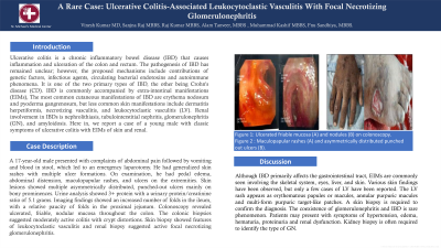
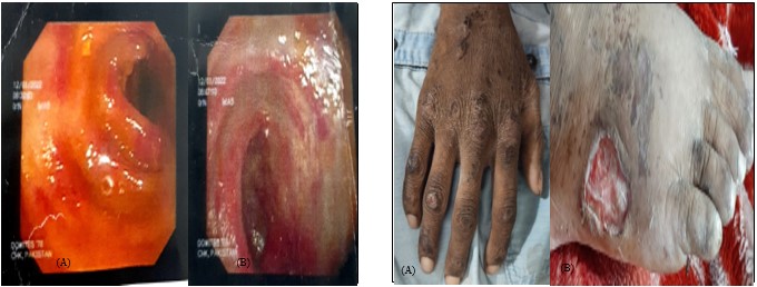
Case Description/Methods: A 17-year-old male presented with complaints of abdominal pain followed by vomiting and blood in stool, which led to an emergency laparotomy. He had generalized skin rashes with multiple ulcer formations. On examination, he had pedal edema, abdominal distension, maculopapular rashes, and ulcers on the extremities. Skin lesions showed multiple asymmetrically distributed, punched-out ulcers mainly on bony prominences. Urine analysis showed 3+ protein with a urinary protein/creatinine ratio of 5.1 grams. Imaging findings showed an increased number of folds in the ileum, with a relative paucity of folds in the proximal jejunum. Colonoscopy revealed ulcerated, friable, nodular mucosa throughout the colon. The colonic biopsies suggested moderately active colitis with crypt distortions. Skin biopsy showed features of leukocytoclastic vasculitis and renal biopsy suggested active focal necrotizing glomerulonephritis.
Discussion: Although IBD primarily affects the gastrointestinal tract, EIMs are commonly seen involving the skeletal system, eyes, liver, and skin. Various skin findings have been observed, but only a few cases of LV have been reported. The LV rash appears as erythematous papules or macules, annular purpuric macules and multi-form purpuric target-like patches. A skin biopsy is required to confirm the diagnosis. The coexistence of glomerulonephritis and IBD is rare phenomenon. Patients may present with symptoms of hypertension, edema, hematuria, proteinuria and renal dysfunction. Kidney biopsy is often required to identify the type of GN.

Disclosures:
Vinesh Kumar, MBBS, MD1, Sanjna Raj, 2, Raj Kumar, MBBS3, Alam Tanveer, MBBS3, Muhammad kashif, MBBS3, Fnu Sandhiya, MBBS4. P4436 - A Rare Case: Ulcerative Colitis-Associated Leukocytoclastic Vasculitis With Focal Necrotizing Glomerulonephritis, ACG 2024 Annual Scientific Meeting Abstracts. Philadelphia, PA: American College of Gastroenterology.
1New York Medical College - Saint Michael's Medical Center, Newark, NJ; 2Bahria University Medical and Dental College, Karachi, Sindh, Pakistan; 3Dow University of Health Sciences, Karachi, Sindh, Pakistan; 4Chandka Medical College, Sukkur, Sindh, Pakistan
Introduction: Ulcerative colitis is a chronic inflammatory bowel disease (IBD) that causes inflammation and ulceration of the colon and rectum. The pathogenesis of IBD has remained unclear; however, the proposed mechanisms include contributions of genetic factors, infectious agents, circulating bacterial endotoxins and autoimmune phenomena. It is one of the two primary types of IBD, the other being Crohn's disease (CD). IBD is commonly accompanied by extra-intestinal manifestations (EIMs), The most common cutaneous manifestations of IBD are erythema nodosum and pyoderma gangrenosum, but less common skin manifestations include dermatitis herpetiformis, necrotizing vasculitis, and leukocytoclastic vasculitis (LV). Renal involvement in IBDs is nephrolithiasis, tubulointerstitial nephritis, glomerulonephritis (GN), and amyloidosis. Here in, we report a case of a young male with classic symptoms of ulcerative colitis with EIMs of skin and renal.
Case Description/Methods: A 17-year-old male presented with complaints of abdominal pain followed by vomiting and blood in stool, which led to an emergency laparotomy. He had generalized skin rashes with multiple ulcer formations. On examination, he had pedal edema, abdominal distension, maculopapular rashes, and ulcers on the extremities. Skin lesions showed multiple asymmetrically distributed, punched-out ulcers mainly on bony prominences. Urine analysis showed 3+ protein with a urinary protein/creatinine ratio of 5.1 grams. Imaging findings showed an increased number of folds in the ileum, with a relative paucity of folds in the proximal jejunum. Colonoscopy revealed ulcerated, friable, nodular mucosa throughout the colon. The colonic biopsies suggested moderately active colitis with crypt distortions. Skin biopsy showed features of leukocytoclastic vasculitis and renal biopsy suggested active focal necrotizing glomerulonephritis.
Discussion: Although IBD primarily affects the gastrointestinal tract, EIMs are commonly seen involving the skeletal system, eyes, liver, and skin. Various skin findings have been observed, but only a few cases of LV have been reported. The LV rash appears as erythematous papules or macules, annular purpuric macules and multi-form purpuric target-like patches. A skin biopsy is required to confirm the diagnosis. The coexistence of glomerulonephritis and IBD is rare phenomenon. Patients may present with symptoms of hypertension, edema, hematuria, proteinuria and renal dysfunction. Kidney biopsy is often required to identify the type of GN.

Figure: Figure 1: Ulcerated friable mucosa (A) and nodules (B) on colonoscopy
Figure 2 : Maculopapular rashes (A) and asymmetrically distributed punched out ulcers (B).
Figure 2 : Maculopapular rashes (A) and asymmetrically distributed punched out ulcers (B).
Disclosures:
Vinesh Kumar indicated no relevant financial relationships.
Sanjna Raj indicated no relevant financial relationships.
Raj Kumar indicated no relevant financial relationships.
Alam Tanveer indicated no relevant financial relationships.
Muhammad kashif indicated no relevant financial relationships.
Fnu Sandhiya indicated no relevant financial relationships.
Vinesh Kumar, MBBS, MD1, Sanjna Raj, 2, Raj Kumar, MBBS3, Alam Tanveer, MBBS3, Muhammad kashif, MBBS3, Fnu Sandhiya, MBBS4. P4436 - A Rare Case: Ulcerative Colitis-Associated Leukocytoclastic Vasculitis With Focal Necrotizing Glomerulonephritis, ACG 2024 Annual Scientific Meeting Abstracts. Philadelphia, PA: American College of Gastroenterology.
