Tuesday Poster Session
Category: GI Bleeding
P4251 - Diffuse Gastrointestinal Amyloidosis in a Patient With Multiple Myeloma
Tuesday, October 29, 2024
10:30 AM - 4:00 PM ET
Location: Exhibit Hall E

Has Audio
.jpg)
Shayan Amini, MD
Houston Methodist-Weill Cornell Graduate School of Medical Sciences
Houston, TX
Presenting Author(s)
Shayan Amini, MD1, Usman Ansari, DO2, Mary R. Schwartz, MD2, Rachel Schiesser, MD3
1Houston Methodist-Weill Cornell Graduate School of Medical Sciences, Houston, TX; 2Houston Methodist Hospital, Houston, TX; 3Underwood Center for Digestive Disorders, Houston Methodist Hospital, Houston, TX
Introduction: Gastrointestinal amyloidosis (GIA) is a rare phenomenon that can be present as both localized and systemic amyloidosis. We report a case of GIA in a young man who presented with weight loss and hematochezia.
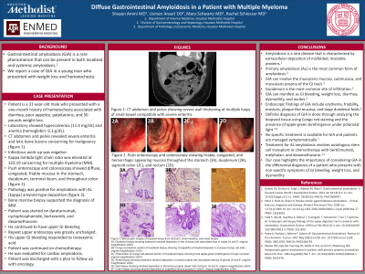
Case Description/Methods: The patient is a 31-year-old male who presented with a one-month history of hematochezia associated with diarrhea, poor appetite, palpitations, and 50-pound weight loss. Laboratory evaluation on presentation showed hypercalcemia (11.4 mg/dL) and anemia with hemoglobin of 6.3 g/dL. A CT abdomen and pelvis revealed severe enteritis and lytic bone lesions concerning for malignancy (Figure 1). An infectious work-up was negative. Serum kappa:lambda light chain ratio was elevated at 323.19 concerning for multiple myeloma (MM). Subsequent push enteroscopy and colonoscopy showed diffuse, congested, friable mucosa in the stomach, duodenum, terminal ileum, and throughout the colon (Figure 2). Biopsies were obtained and pathology demonstrated amyloid deposition at each site, typed as AL (kappa) amyloid (Figure 3). Bone marrow biopsy supported the diagnosis of MM. The patient was started on daratumumab, cyclophosphamide, bortezomib, and dexamethasone. Unfortunately, he continued to have upper gastrointestinal (GI) bleeding and findings at repeat upper endoscopy were unchanged. Treatment with tranexamic was successful in stabilizing his GI bleeding. He continued chemotherapy, underwent evaluation for cardiac amyloidosis, and was discharged with a plan to follow up with oncology.
Discussion: Amyloidosis is a rare disease that is characterized by extracellular deposition of misfolded, insoluble proteins. Primary amyloidosis (AL) is the most common form of amyloidosis and can involve the mucosa, muscularis mucosa, submucosa, and muscularis propria of the GI tract. GIA can manifest as GI bleeding, weight loss, diarrhea, dysmotility, and ascites. Endoscopic findings of GIA include erythema, friability, erosions, plaque-like mucosa, and large duodenal folds. A diagnosis of GIA is confirmed using Congo red staining and amyloid subtyping can be performed on paraffin-embeded biopsies. No specific treatment is available for GIA and patients are managed symptomatically. Treatment for AL amyloidosis involves autologous stem cell transplant or chemotherapy with bertilimumab, melphalan, and dexamethasone. Our case highlights the importance of considering GIA in the differential diagnosis of a patient who presents with non-specific symptoms of GI bleeding, weight loss, and dysmotility.

Disclosures:
Shayan Amini, MD1, Usman Ansari, DO2, Mary R. Schwartz, MD2, Rachel Schiesser, MD3. P4251 - Diffuse Gastrointestinal Amyloidosis in a Patient With Multiple Myeloma, ACG 2024 Annual Scientific Meeting Abstracts. Philadelphia, PA: American College of Gastroenterology.
1Houston Methodist-Weill Cornell Graduate School of Medical Sciences, Houston, TX; 2Houston Methodist Hospital, Houston, TX; 3Underwood Center for Digestive Disorders, Houston Methodist Hospital, Houston, TX
Introduction: Gastrointestinal amyloidosis (GIA) is a rare phenomenon that can be present as both localized and systemic amyloidosis. We report a case of GIA in a young man who presented with weight loss and hematochezia.
Case Description/Methods: The patient is a 31-year-old male who presented with a one-month history of hematochezia associated with diarrhea, poor appetite, palpitations, and 50-pound weight loss. Laboratory evaluation on presentation showed hypercalcemia (11.4 mg/dL) and anemia with hemoglobin of 6.3 g/dL. A CT abdomen and pelvis revealed severe enteritis and lytic bone lesions concerning for malignancy (Figure 1). An infectious work-up was negative. Serum kappa:lambda light chain ratio was elevated at 323.19 concerning for multiple myeloma (MM). Subsequent push enteroscopy and colonoscopy showed diffuse, congested, friable mucosa in the stomach, duodenum, terminal ileum, and throughout the colon (Figure 2). Biopsies were obtained and pathology demonstrated amyloid deposition at each site, typed as AL (kappa) amyloid (Figure 3). Bone marrow biopsy supported the diagnosis of MM. The patient was started on daratumumab, cyclophosphamide, bortezomib, and dexamethasone. Unfortunately, he continued to have upper gastrointestinal (GI) bleeding and findings at repeat upper endoscopy were unchanged. Treatment with tranexamic was successful in stabilizing his GI bleeding. He continued chemotherapy, underwent evaluation for cardiac amyloidosis, and was discharged with a plan to follow up with oncology.
Discussion: Amyloidosis is a rare disease that is characterized by extracellular deposition of misfolded, insoluble proteins. Primary amyloidosis (AL) is the most common form of amyloidosis and can involve the mucosa, muscularis mucosa, submucosa, and muscularis propria of the GI tract. GIA can manifest as GI bleeding, weight loss, diarrhea, dysmotility, and ascites. Endoscopic findings of GIA include erythema, friability, erosions, plaque-like mucosa, and large duodenal folds. A diagnosis of GIA is confirmed using Congo red staining and amyloid subtyping can be performed on paraffin-embeded biopsies. No specific treatment is available for GIA and patients are managed symptomatically. Treatment for AL amyloidosis involves autologous stem cell transplant or chemotherapy with bertilimumab, melphalan, and dexamethasone. Our case highlights the importance of considering GIA in the differential diagnosis of a patient who presents with non-specific symptoms of GI bleeding, weight loss, and dysmotility.

Figure: Figure 1: CT abdomen and pelvis showing severe wall thickening of multiple loops of small bowel compatible with severe enteritis.
Figure 2: Push enteroscopy and colonoscopy showing friable, congested, and hemorrhagic appearing mucosa throughout the stomach, duodenum, sigmoid colon, and rectum.
Figure 3: Microscopic images of biopsied tissue from stomach, small intestine, and colon biopsy
3A) Duodenal biopsy showing extensive amyloid deposition in the mucosa with associated loss of crypts (H and E, original magnification x200)
3B) Congo red stained section of duodenal biopsy showing Congophilia of amyloid deposits in mucosa (Congo red stain, original magnification x100)
3C) Polarization of Congo red-stained section of duodenal biopsy showing focal apple-green birefringence (Congo red stain, original magnification x200)
3D) Antral biopsy showing extensive amyloid deposition in lamina propria with decreased density of glands (H and E, original magnification x200)
3E) Ileal biopsy showing amyloid deposition in the lamina propria (lower half of photo). (H and E, original magnification x200)
3F) Colon biopsy showing amyloid deposition in superficial lamina propria (H and E, original magnification x200)
Disclosures:
Shayan Amini indicated no relevant financial relationships.
Usman Ansari indicated no relevant financial relationships.
Mary Schwartz indicated no relevant financial relationships.
Rachel Schiesser indicated no relevant financial relationships.
Shayan Amini, MD1, Usman Ansari, DO2, Mary R. Schwartz, MD2, Rachel Schiesser, MD3. P4251 - Diffuse Gastrointestinal Amyloidosis in a Patient With Multiple Myeloma, ACG 2024 Annual Scientific Meeting Abstracts. Philadelphia, PA: American College of Gastroenterology.
