Monday Poster Session
Category: Colon
P1990 - History Matters: A Rare Case of Rectosigmoid Endometriosis Disguised as Colorectal Malignancy
Monday, October 28, 2024
10:30 AM - 4:00 PM ET
Location: Exhibit Hall E

Has Audio

Nguyen Pham, MD
Ronald Reagan Hospital
Los Angeles, CA
Presenting Author(s)
Award: Presidential Poster Award
Nguyen Pham, MD1, Kirstin H. Peters, MD2, Janice Oh, MD, MS3, Kevin A.. Ghassemi, MD4
1Ronald Reagan Hospital, Los Angeles, CA; 2University of California Los Angeles Health, Los Angeles, CA; 3UCLA, Los Angeles, CA; 4David Geffen School of Medicine at UCLA, Los Angeles, CA
Introduction: Endometriosis involves extrauterine growth of endometrial tissue. It can lead to nonspecific gastrointestinal (GI) symptoms including abdominal pain, and constipation but severe obstruction is rare. We present a rare case of rectosigmoid endometriosis causing large bowel obstruction in a female patient with a history of infertility.
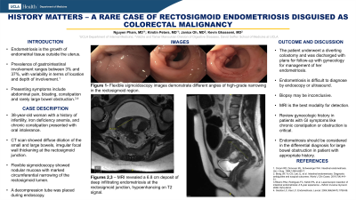
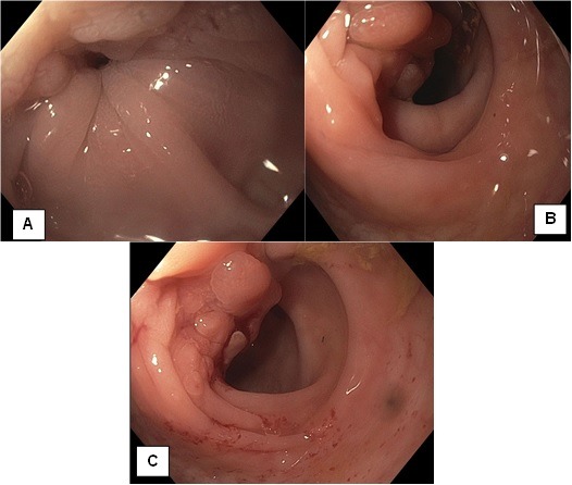
Case Description/Methods: A 38-year-old female with a history of infertility, iron deficiency anemia, and chronic constipation, presented with oral intolerance. One year prior, a computed tomography (CT) scan of the abdomen and pelvis demonstrated left ovarian cysts, bilateral hydrosalpinx, and uterine thickening. Repeat CT on admission revealed a diffusely dilated small and large bowel with irregular focal wall thickening at the rectosigmoid junction. An urgent flexible sigmoidoscopy demonstrated nodular mucosa with marked circumferential narrowing of the rectosigmoid lumen and a decompression tube was placed (Exhibit 1A-C). Magnetic Resonance Imaging of the abdomen and pelvis revealed a large, thick deposit of deep infiltrating endometriosis involving 6.8 cm of the rectosigmoid. She ultimately underwent a diverting colostomy and was discharged with close gynecology follow up for further management of her endometriosis.
Discussion: Gastrointestinal endometriosis occurs in 3-37% of patients, and most commonly affects the rectosigmoid area as seen in our patient. In this case, an important clue to intestinal endometriosis was the temporal pattern between constipation and infertility in her history. Diagnosing endometriosis of the GI tract can be challenging as it might not be consistently detected by imaging or endoscopic evaluation. Often, surgical exploration is needed. If detected endoscopically, endometriosis can appear as nodular growth, mimicking adenocarcinoma. Endoscopic biopsy might be non-diagnostic as endometriosis will not always infiltrate the mucosal layer. MRI can be helpful to identify endometriosis, as it will appear as a hypointense signal on T2-weighted imaging.
This case highlights the importance of reviewing gynecologic history in patients who present with GI symptoms. In the correct context, endometriosis should be included in the differential for chronic constipation and acute large bowel obstruction. When suspicion is high, MRI and laparoscopy are the best diagnostics tools. Timely evaluation of endometriosis is crucial to prevent severe GI complications.

Disclosures:
Nguyen Pham, MD1, Kirstin H. Peters, MD2, Janice Oh, MD, MS3, Kevin A.. Ghassemi, MD4. P1990 - History Matters: A Rare Case of Rectosigmoid Endometriosis Disguised as Colorectal Malignancy, ACG 2024 Annual Scientific Meeting Abstracts. Philadelphia, PA: American College of Gastroenterology.
Nguyen Pham, MD1, Kirstin H. Peters, MD2, Janice Oh, MD, MS3, Kevin A.. Ghassemi, MD4
1Ronald Reagan Hospital, Los Angeles, CA; 2University of California Los Angeles Health, Los Angeles, CA; 3UCLA, Los Angeles, CA; 4David Geffen School of Medicine at UCLA, Los Angeles, CA
Introduction: Endometriosis involves extrauterine growth of endometrial tissue. It can lead to nonspecific gastrointestinal (GI) symptoms including abdominal pain, and constipation but severe obstruction is rare. We present a rare case of rectosigmoid endometriosis causing large bowel obstruction in a female patient with a history of infertility.
Case Description/Methods: A 38-year-old female with a history of infertility, iron deficiency anemia, and chronic constipation, presented with oral intolerance. One year prior, a computed tomography (CT) scan of the abdomen and pelvis demonstrated left ovarian cysts, bilateral hydrosalpinx, and uterine thickening. Repeat CT on admission revealed a diffusely dilated small and large bowel with irregular focal wall thickening at the rectosigmoid junction. An urgent flexible sigmoidoscopy demonstrated nodular mucosa with marked circumferential narrowing of the rectosigmoid lumen and a decompression tube was placed (Exhibit 1A-C). Magnetic Resonance Imaging of the abdomen and pelvis revealed a large, thick deposit of deep infiltrating endometriosis involving 6.8 cm of the rectosigmoid. She ultimately underwent a diverting colostomy and was discharged with close gynecology follow up for further management of her endometriosis.
Discussion: Gastrointestinal endometriosis occurs in 3-37% of patients, and most commonly affects the rectosigmoid area as seen in our patient. In this case, an important clue to intestinal endometriosis was the temporal pattern between constipation and infertility in her history. Diagnosing endometriosis of the GI tract can be challenging as it might not be consistently detected by imaging or endoscopic evaluation. Often, surgical exploration is needed. If detected endoscopically, endometriosis can appear as nodular growth, mimicking adenocarcinoma. Endoscopic biopsy might be non-diagnostic as endometriosis will not always infiltrate the mucosal layer. MRI can be helpful to identify endometriosis, as it will appear as a hypointense signal on T2-weighted imaging.
This case highlights the importance of reviewing gynecologic history in patients who present with GI symptoms. In the correct context, endometriosis should be included in the differential for chronic constipation and acute large bowel obstruction. When suspicion is high, MRI and laparoscopy are the best diagnostics tools. Timely evaluation of endometriosis is crucial to prevent severe GI complications.

Figure: Exhibit 1: A, B, C - Flexible sigmoidoscopy images demonstrate different angles of a large irregular infiltrative and obstructive proximal rectal mass.
Disclosures:
Nguyen Pham indicated no relevant financial relationships.
Kirstin Peters indicated no relevant financial relationships.
Janice Oh indicated no relevant financial relationships.
Kevin Ghassemi indicated no relevant financial relationships.
Nguyen Pham, MD1, Kirstin H. Peters, MD2, Janice Oh, MD, MS3, Kevin A.. Ghassemi, MD4. P1990 - History Matters: A Rare Case of Rectosigmoid Endometriosis Disguised as Colorectal Malignancy, ACG 2024 Annual Scientific Meeting Abstracts. Philadelphia, PA: American College of Gastroenterology.

