Monday Poster Session
Category: Colon
P1997 - Metastatic Merkel Cell Carcinoma of Unknown Primary Presenting as an Exophytic Colon Mass
Monday, October 28, 2024
10:30 AM - 4:00 PM ET
Location: Exhibit Hall E

Has Audio
- KL
Krystal Y. Lai, MD
University of Texas Southwestern
Dallas, TX
Presenting Author(s)
Award: Presidential Poster Award
Krystal Y. Lai, MD1, Purva Gopal, MD1, Ariel W. Aday, MD2
1University of Texas Southwestern, Dallas, TX; 2UT Southwestern, Dallas, TX
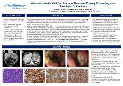
Introduction: Merkel cell carcinoma (MCC) is a rare neuroendocrine tumor of the skin associated with high rates of recurrence and metastasis. Common sites of metastasis are, in order of frequency, distant lymph nodes, skin, liver, bone, pancreas, lung, and brain. Metastasis to the gastrointestinal tract is rare and most of these documented cases involve the anal canal, stomach and small intestine. Less common is metastasis to the colon.
Case Description/Methods: A 67-year-old Hispanic male with no known past medical history presented to the emergency department with 8 days of left lower quadrant pain, hematochezia, nausea and weight loss. On admission, the patient was tachycardic, and hemoglobin was 8.6 g/dL. He had no prior laboratory values for comparison, no known history of cancer, and no reported skin lesions. The computed tomography (CT) of the abdomen and pelvis showed a 14.8 cm x 12.9 cm x 14.4 cm exophytic mass in the transverse colon with extension into the stomach, spleen, and small bowel. Numerous peritoneal implants, an enlarged right external iliac lymph node, and bilateral adrenal nodules were also noted. The CT of the chest was only positive for parenchymal nodules measuring less than 3 mm. Upper endoscopy revealed a large, ulcerated mass on the lesser curvature of the gastric body with an adjacent medium-sized, ulcerated mass on the greater curvature of the gastric body. Colonoscopy revealed an 8 cm infiltrative, ulcerated, partially obstructing mass in the transverse colon. Biopsies of gastric and colonic masses were obtained. The neoplastic cells were positive for cytokeratin AE1/AE3, synaptophysin, and chromogranin and negative for CD45 and CDX2. Additionally, CK20 stained in a perinuclear, dot-like positivity consistent with metastatic Merkel cell carcinoma. Ki-67 stain highlighted 90% of neoplastic cells.
Discussion: Here we present a case of Merkel cell carcinoma of unknown primary (MCCUP) with metastases to lymph nodes, adrenal glands, peritoneum, stomach, small bowel, and colon, presenting as a large exophytic colonic mass on imaging. There have been 9 case reports of MCC metastasis to the colon, of which 2 have been within the transverse colon. Most of these cases have occurred in patients with previously known diagnoses of primary MCC skin lesions. Only one prior case of metastatic MCCUP to the colon has been previously documented. Biopsy of the gastrointestinal involvement is essential for diagnosis as presentation may occur without any prior documentation of MCC.

Disclosures:
Krystal Y. Lai, MD1, Purva Gopal, MD1, Ariel W. Aday, MD2. P1997 - Metastatic Merkel Cell Carcinoma of Unknown Primary Presenting as an Exophytic Colon Mass, ACG 2024 Annual Scientific Meeting Abstracts. Philadelphia, PA: American College of Gastroenterology.
Krystal Y. Lai, MD1, Purva Gopal, MD1, Ariel W. Aday, MD2
1University of Texas Southwestern, Dallas, TX; 2UT Southwestern, Dallas, TX
Introduction: Merkel cell carcinoma (MCC) is a rare neuroendocrine tumor of the skin associated with high rates of recurrence and metastasis. Common sites of metastasis are, in order of frequency, distant lymph nodes, skin, liver, bone, pancreas, lung, and brain. Metastasis to the gastrointestinal tract is rare and most of these documented cases involve the anal canal, stomach and small intestine. Less common is metastasis to the colon.
Case Description/Methods: A 67-year-old Hispanic male with no known past medical history presented to the emergency department with 8 days of left lower quadrant pain, hematochezia, nausea and weight loss. On admission, the patient was tachycardic, and hemoglobin was 8.6 g/dL. He had no prior laboratory values for comparison, no known history of cancer, and no reported skin lesions. The computed tomography (CT) of the abdomen and pelvis showed a 14.8 cm x 12.9 cm x 14.4 cm exophytic mass in the transverse colon with extension into the stomach, spleen, and small bowel. Numerous peritoneal implants, an enlarged right external iliac lymph node, and bilateral adrenal nodules were also noted. The CT of the chest was only positive for parenchymal nodules measuring less than 3 mm. Upper endoscopy revealed a large, ulcerated mass on the lesser curvature of the gastric body with an adjacent medium-sized, ulcerated mass on the greater curvature of the gastric body. Colonoscopy revealed an 8 cm infiltrative, ulcerated, partially obstructing mass in the transverse colon. Biopsies of gastric and colonic masses were obtained. The neoplastic cells were positive for cytokeratin AE1/AE3, synaptophysin, and chromogranin and negative for CD45 and CDX2. Additionally, CK20 stained in a perinuclear, dot-like positivity consistent with metastatic Merkel cell carcinoma. Ki-67 stain highlighted 90% of neoplastic cells.
Discussion: Here we present a case of Merkel cell carcinoma of unknown primary (MCCUP) with metastases to lymph nodes, adrenal glands, peritoneum, stomach, small bowel, and colon, presenting as a large exophytic colonic mass on imaging. There have been 9 case reports of MCC metastasis to the colon, of which 2 have been within the transverse colon. Most of these cases have occurred in patients with previously known diagnoses of primary MCC skin lesions. Only one prior case of metastatic MCCUP to the colon has been previously documented. Biopsy of the gastrointestinal involvement is essential for diagnosis as presentation may occur without any prior documentation of MCC.

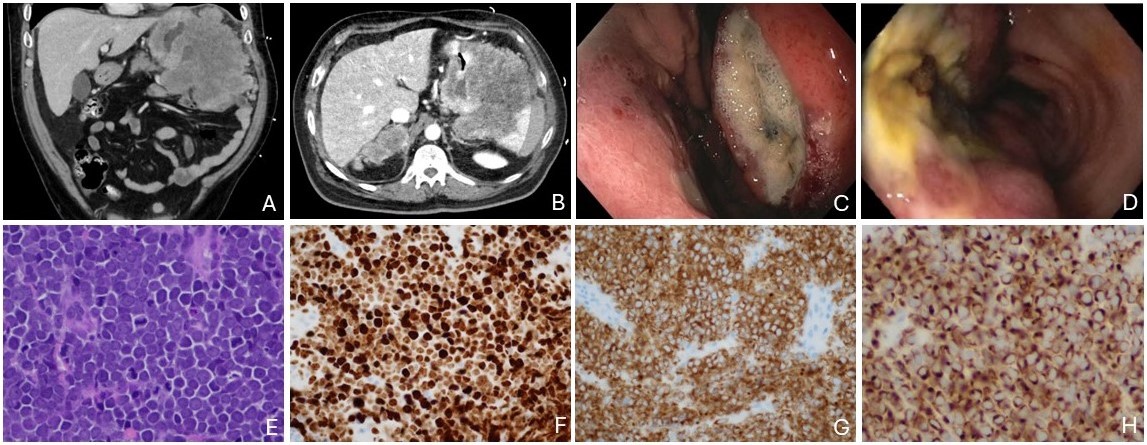
Figure: Figure 1. (A) Coronal and (B) axial planes of CT abdomen and pelvis with 14.8cm x 12.9cm x 14.4cm left upper quadrant mass centered on the transverse colon with extension to adjacent stomach, spleen, and small bowel. (C) Upper endoscopy image of large, ulcerated, invasive mass in gastric body. (D) Colonoscopy image of infiltrative, ulcerated, non-circumferential, partially obstructing 8cm mass in transverse colon. Colon biopsy histology: (E) H&E stain showing cytologic features of the tumor, nuclear molding, salt and pepper chromatin, and several mitotic figures at 60x, (F) ki67 immunohistochemical (IHC) stain highlighting nearly 100% of the neoplastic cells at 40x, (G) synaptophysin IHC stain at 40x, (H) CK20 IHC stain highlighting neoplastic cells with perinuclear dot-like positivity, characteristic of MCC at 60x.
Disclosures:
Krystal Lai indicated no relevant financial relationships.
Purva Gopal indicated no relevant financial relationships.
Ariel Aday indicated no relevant financial relationships.
Krystal Y. Lai, MD1, Purva Gopal, MD1, Ariel W. Aday, MD2. P1997 - Metastatic Merkel Cell Carcinoma of Unknown Primary Presenting as an Exophytic Colon Mass, ACG 2024 Annual Scientific Meeting Abstracts. Philadelphia, PA: American College of Gastroenterology.

