Monday Poster Session
Category: Colon
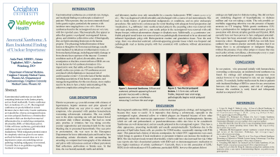
P2004 - Anorectal Xanthoma: A Rare Incidental Finding of Unclear Importance
Monday, October 28, 2024
10:30 AM - 4:00 PM ET
Location: Exhibit Hall E

Has Audio
.jpg)
Amla A. Patel, MBBS
Creighton University
Phoenix, AZ
Presenting Author(s)
Amla A. Patel, MBBS1, Ariana R. Tagliaferri, MD2, Andrew M. Weinberg, DO3
1Creighton University, Phoenix, AZ; 2Creighton University Medical Center, Phoenix, AZ; 3Creighton University School of Medicine, Scottsdale, AZ
Introduction: Gastrointestinal xanthomas are rare lipid-containing histiocytes in the lamina propria, and are found incidentally. Gastric xanthomas have an incidence of 0.2-0.8%. Rectosigmoid xanthomas are poorly defined in current literature due to their rarity. Gastric xanthomas are typically flat, whereas rectosigmoid lesions are more polypoid. Both have a whitish-yellow coloration which can develop from mucosal inflammation due to underlying constipation, diverticulitis, IBD, or Helicobacter pylori. Unlike soft tissue xanthomas, gastrointestinal xanthomas are not correlated with dyslipidemia. We present a patient found to have flat rectal xanthomas near the anal verge.
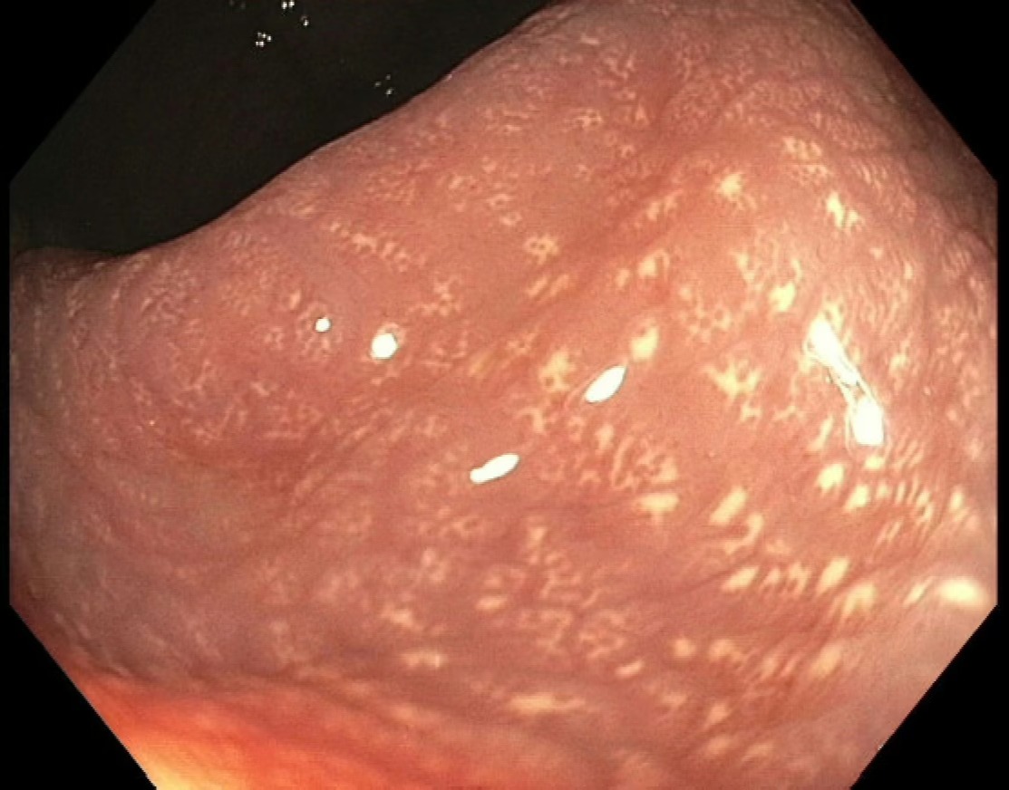
Case Description/Methods: A 40 year-old woman with PMH of HTN and hepatic steatosis presented to GI clinic for evaluation of painless hematochezia. She had a history of uncomplicated diverticulitis one year prior, had soft regular bowel movements without straining and was taking Metamucil daily. She did not have a history of known hemorrhoids. A colonoscopy with adequate visualization revealed a 4 mm sessile-serrated polyp without adenomatous changes on pathology, two ulcerated and friable hyperplastic polyps of the rectum without adenomatous changes, and abnormal appearing, scattered and flat yellowish-appearing granulation tissue near the anal verge (Figure 1). Biopsies of this revealed lamina propria with foci consistent with xanthoma without adenomatous changes.
Discussion: Our case is unique in that not only was her lesion flat, which is more typical of gastric xanthomas, but her lesions were also closer to the anal verge rather than at the rectosigmoid junction. This is thus the first case of anorectal xanthomas described in current literature. As this patient’s diverticular disease was restricted to the descending colon, it is not plausible to be causative of her anorectal xanthomas. In the absence of chronic constipation or hemorrhoids, ultimately the underlying etiology is unknown. While gastric xanthomas have a predisposition for malignant transformation due to association with chronic atrophic gastritis and H. pylori, the malignant potential of rectosigmoid xanthomas is ultimately unknown. Further studies are indicated to assess for other risk factors, etiologies and management following incidental identification of rectosigmoid xanthomas.

Disclosures:
Amla A. Patel, MBBS1, Ariana R. Tagliaferri, MD2, Andrew M. Weinberg, DO3. P2004 - Anorectal Xanthoma: A Rare Incidental Finding of Unclear Importance, ACG 2024 Annual Scientific Meeting Abstracts. Philadelphia, PA: American College of Gastroenterology.
1Creighton University, Phoenix, AZ; 2Creighton University Medical Center, Phoenix, AZ; 3Creighton University School of Medicine, Scottsdale, AZ
Introduction: Gastrointestinal xanthomas are rare lipid-containing histiocytes in the lamina propria, and are found incidentally. Gastric xanthomas have an incidence of 0.2-0.8%. Rectosigmoid xanthomas are poorly defined in current literature due to their rarity. Gastric xanthomas are typically flat, whereas rectosigmoid lesions are more polypoid. Both have a whitish-yellow coloration which can develop from mucosal inflammation due to underlying constipation, diverticulitis, IBD, or Helicobacter pylori. Unlike soft tissue xanthomas, gastrointestinal xanthomas are not correlated with dyslipidemia. We present a patient found to have flat rectal xanthomas near the anal verge.
Case Description/Methods: A 40 year-old woman with PMH of HTN and hepatic steatosis presented to GI clinic for evaluation of painless hematochezia. She had a history of uncomplicated diverticulitis one year prior, had soft regular bowel movements without straining and was taking Metamucil daily. She did not have a history of known hemorrhoids. A colonoscopy with adequate visualization revealed a 4 mm sessile-serrated polyp without adenomatous changes on pathology, two ulcerated and friable hyperplastic polyps of the rectum without adenomatous changes, and abnormal appearing, scattered and flat yellowish-appearing granulation tissue near the anal verge (Figure 1). Biopsies of this revealed lamina propria with foci consistent with xanthoma without adenomatous changes.
Discussion: Our case is unique in that not only was her lesion flat, which is more typical of gastric xanthomas, but her lesions were also closer to the anal verge rather than at the rectosigmoid junction. This is thus the first case of anorectal xanthomas described in current literature. As this patient’s diverticular disease was restricted to the descending colon, it is not plausible to be causative of her anorectal xanthomas. In the absence of chronic constipation or hemorrhoids, ultimately the underlying etiology is unknown. While gastric xanthomas have a predisposition for malignant transformation due to association with chronic atrophic gastritis and H. pylori, the malignant potential of rectosigmoid xanthomas is ultimately unknown. Further studies are indicated to assess for other risk factors, etiologies and management following incidental identification of rectosigmoid xanthomas.

Figure: Figure 1. Anorectal Xanthoma. Diffuse and scattered, yellowish-appearing flat and granular mucosa with a shining appearance, seen on retroflexion, measuring 2 cm from the anal verge.
Disclosures:
Amla Patel indicated no relevant financial relationships.
Ariana Tagliaferri indicated no relevant financial relationships.
Andrew Weinberg indicated no relevant financial relationships.
Amla A. Patel, MBBS1, Ariana R. Tagliaferri, MD2, Andrew M. Weinberg, DO3. P2004 - Anorectal Xanthoma: A Rare Incidental Finding of Unclear Importance, ACG 2024 Annual Scientific Meeting Abstracts. Philadelphia, PA: American College of Gastroenterology.
