Sunday Poster Session
Category: Biliary/Pancreas
P0099 - Maybe This Horse is a Zebra - Pancreatitis or Omental Infarct?
Sunday, October 27, 2024
3:30 PM - 7:00 PM ET
Location: Exhibit Hall E

Has Audio
- AS
Andrea Schneider, DO
HonorHealth
Phoenix, AZ
Presenting Author(s)
Andrea Schneider, DO1, Gavin Levinthal, MD2
1HonorHealth, Phoenix, AZ; 2HonorHealth, Scottsdale, AZ
Introduction: Omental infarction occurs due to compromised omental blood supply. Causes include torsion, thrombosis, trauma, obesity, prior surgeries, or may be unexplained. Omental infarction is a rare condition with approximately 400 cases documented in the literature. We are aware of only one other occurrence of a patient with pancreatitis and omental infarction. We share a similar case, but our patient also had a portal vein thrombosis.
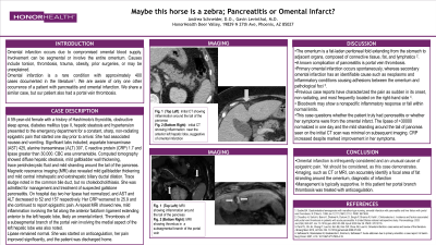
Case Description/Methods: A 59-year-old female with a history of obstructive sleep apnea, diabetes mellitus type II, hepatic steatosis and hypertension presented to the emergency department for a constant, sharp, non-radiating epigastric pain. Significant labs included; aspartate transaminase 425, alanine transaminase 307, C reactive protein 1.7 and lipase greater than 30,000. Computed tomography showed diffuse hepatic steatosis, mild gallbladder wall thickening, trace pericholecystic fluid and mild stranding around the tail of the pancreas. Magnetic resonance imaging also revealed mild gallbladder thickening and mild central intrahepatic and extrahepatic biliary ductal dilation. Trace sludge noted in the common bile duct, but no choledocholithiasis. She was admitted for management and treatment of suspected gallstone pancreatitis. On hospital day two her lipase had normalized and liver enzymes improved. Her CRP worsened to 25.9 and she continued to report epigastric pain. A repeat MRI showed new, mild inflammation involving the fat along the anterior falciform ligament extending anterior to the left hepatic lobe, likely an omental infarct. Thrombosis of a subsegmental branch of the portal vein involving the medial aspect of the left hepatic lobe was also noted. Lipase remained normal. She was started on anticoagulation, her pain improved significantly, and the patient was discharged home.
Discussion: The omentum is a fat-laden peritoneal fold extending from the stomach to adjacent organs, composed of connective tissue, fat, and lymphatics. Primary omental infarction occurs spontaneously, whereas secondary omental infarction has an identifiable cause such as inflammatory conditions causing adhesions between the omentum and pathological foci. This case questions whether the patient had pancreatitis or whether her symptoms were from the omental infarct. The lipase of >35000 normalized in one day and the mild stranding around the tail of pancreas seen on the initial CT scan was minimal on subsequent imaging. CRP increased despite marked improvement in her symptoms.

Disclosures:
Andrea Schneider, DO1, Gavin Levinthal, MD2. P0099 - Maybe This Horse is a Zebra - Pancreatitis or Omental Infarct?, ACG 2024 Annual Scientific Meeting Abstracts. Philadelphia, PA: American College of Gastroenterology.
1HonorHealth, Phoenix, AZ; 2HonorHealth, Scottsdale, AZ
Introduction: Omental infarction occurs due to compromised omental blood supply. Causes include torsion, thrombosis, trauma, obesity, prior surgeries, or may be unexplained. Omental infarction is a rare condition with approximately 400 cases documented in the literature. We are aware of only one other occurrence of a patient with pancreatitis and omental infarction. We share a similar case, but our patient also had a portal vein thrombosis.
Case Description/Methods: A 59-year-old female with a history of obstructive sleep apnea, diabetes mellitus type II, hepatic steatosis and hypertension presented to the emergency department for a constant, sharp, non-radiating epigastric pain. Significant labs included; aspartate transaminase 425, alanine transaminase 307, C reactive protein 1.7 and lipase greater than 30,000. Computed tomography showed diffuse hepatic steatosis, mild gallbladder wall thickening, trace pericholecystic fluid and mild stranding around the tail of the pancreas. Magnetic resonance imaging also revealed mild gallbladder thickening and mild central intrahepatic and extrahepatic biliary ductal dilation. Trace sludge noted in the common bile duct, but no choledocholithiasis. She was admitted for management and treatment of suspected gallstone pancreatitis. On hospital day two her lipase had normalized and liver enzymes improved. Her CRP worsened to 25.9 and she continued to report epigastric pain. A repeat MRI showed new, mild inflammation involving the fat along the anterior falciform ligament extending anterior to the left hepatic lobe, likely an omental infarct. Thrombosis of a subsegmental branch of the portal vein involving the medial aspect of the left hepatic lobe was also noted. Lipase remained normal. She was started on anticoagulation, her pain improved significantly, and the patient was discharged home.
Discussion: The omentum is a fat-laden peritoneal fold extending from the stomach to adjacent organs, composed of connective tissue, fat, and lymphatics. Primary omental infarction occurs spontaneously, whereas secondary omental infarction has an identifiable cause such as inflammatory conditions causing adhesions between the omentum and pathological foci. This case questions whether the patient had pancreatitis or whether her symptoms were from the omental infarct. The lipase of >35000 normalized in one day and the mild stranding around the tail of pancreas seen on the initial CT scan was minimal on subsequent imaging. CRP increased despite marked improvement in her symptoms.

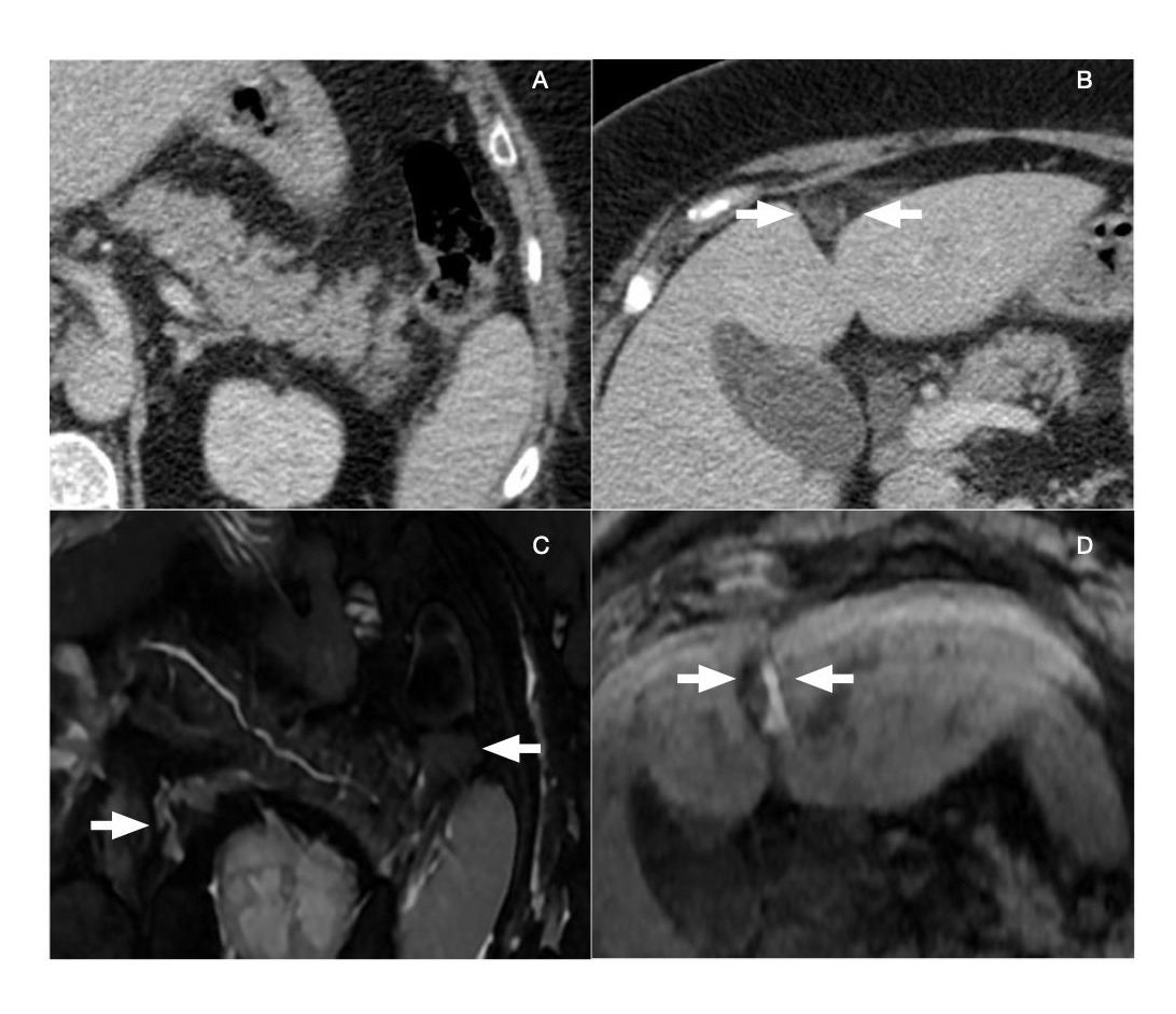
Figure: A CT showing inflammation around the tail of the pancreas
B CT showing inflammation near the anterior left hepatic lobe, suggestive of omental infarction
C MRI showing inflammation around the tail of the pancreas
D MRI showing thrombus in a subsegmental branch of the portal vein
B CT showing inflammation near the anterior left hepatic lobe, suggestive of omental infarction
C MRI showing inflammation around the tail of the pancreas
D MRI showing thrombus in a subsegmental branch of the portal vein
Disclosures:
Andrea Schneider indicated no relevant financial relationships.
Gavin Levinthal indicated no relevant financial relationships.
Andrea Schneider, DO1, Gavin Levinthal, MD2. P0099 - Maybe This Horse is a Zebra - Pancreatitis or Omental Infarct?, ACG 2024 Annual Scientific Meeting Abstracts. Philadelphia, PA: American College of Gastroenterology.
