Sunday Poster Session
Category: Biliary/Pancreas
P0102 - Exploring the Anomalies: A Case of Type Va Duplication of Extrahepatic Biliary Ducts Identified on MRCP
Sunday, October 27, 2024
3:30 PM - 7:00 PM ET
Location: Exhibit Hall E

- WL
Winghang Lau, MD
Nassau University Medical Center
East Meadow, NY
Presenting Author(s)
Raj Mohan Ram Mohan, MD, Winghang Lau, MD, Sai Reshma Magam, MD, Sai Greeshma Magam, MD, Melvin Joy, MD, Dilman Natt, MD, Abhishek Tadikonda, MD, Leeza Pannikodu, MD, Jiten Desai, MD, Paul Mustacchia, MD, MBA
Nassau University Medical Center, East Meadow, NY
Introduction: Duplication of the extrahepatic biliary duct is a rare congenital anomaly. The widely accepted classification of DCBD is the modified version proposed by Choi et al. It describes 5 different types of DCBD, with type Va being one of the rarest forms. Previous cases had reported associations between double common bile duct (CBD) and pancreatitis, cholangitis, and cholecystitis. Here, we report a case of a type Va duplication of bile duct that was incidentally found on an MRCP in a patient undergoing surveillance of a pancreatic lesion.
Case Description/Methods: A 55-year-old female with a past medical history of non-alcoholic hepatic steatosis, transaminitis, gastroesophageal reflux disorder, and pancreatic lesion was followed up in the clinic pancreatic lesion monitoring. The pancreatic lesion was incidentally found in January 2022 after a repeat computed tomography (CT) scan of the abdominal/pelvis showed a 1.5x1.5cm hypodense lesion within the tail of the pancreas. The patient had no complaints and the physical exam was benign. A magnetic resonance imaging (MRI) performed later confirmed the pancreatic lesion. Subsequently, she underwent an endoscopic ultrasound for a potential biopsy of the lesion, and a dual biliary duct joined at the pancreatic head. She was scheduled for a routine MRCP for further evaluation.
Lab results indicated hemoglobin at 12.8 gm/dl, leukocyte count at 7,050/mm3, platelets at 375,000/mm3, total bilirubin at 0.7 mg/dl, AST at 39 U/L, ALT at 54 U/L, and ALP at 155 U/L. MRCP revealed an extrahepatic biliary ductal structure, with two ducts converging in the lower part of the pancreatic head, typical of a type Va variant. Motion artifacts proved difficult to identify whether the anatomy represented a long cystic duct with a low insertion or a very low common bile duct bifurcation. The pancreatic lesion hadn't changed in size from images taken 2 years prior, not requiring medical intervention.
Discussion: We report a case of two long extrahepatic bile ducts joining distally to form one common bile duct with a long cystic duct joining the right extrahepatic duct at a low insertion. Embryogenesis of the hepatobiliary system occurs in the 3rd week of gestation. DCBD has been postulated to be a byproduct of multiple mechanisms, such as random subdivision of hepatic diverticulum, defect in the recanalization process, and disruption of development leading to the persistence of an extrahepatic accessory duct that later regresses to form a single common bile duct.

Disclosures:
Raj Mohan Ram Mohan, MD, Winghang Lau, MD, Sai Reshma Magam, MD, Sai Greeshma Magam, MD, Melvin Joy, MD, Dilman Natt, MD, Abhishek Tadikonda, MD, Leeza Pannikodu, MD, Jiten Desai, MD, Paul Mustacchia, MD, MBA. P0102 - Exploring the Anomalies: A Case of Type Va Duplication of Extrahepatic Biliary Ducts Identified on MRCP, ACG 2024 Annual Scientific Meeting Abstracts. Philadelphia, PA: American College of Gastroenterology.
Nassau University Medical Center, East Meadow, NY
Introduction: Duplication of the extrahepatic biliary duct is a rare congenital anomaly. The widely accepted classification of DCBD is the modified version proposed by Choi et al. It describes 5 different types of DCBD, with type Va being one of the rarest forms. Previous cases had reported associations between double common bile duct (CBD) and pancreatitis, cholangitis, and cholecystitis. Here, we report a case of a type Va duplication of bile duct that was incidentally found on an MRCP in a patient undergoing surveillance of a pancreatic lesion.
Case Description/Methods: A 55-year-old female with a past medical history of non-alcoholic hepatic steatosis, transaminitis, gastroesophageal reflux disorder, and pancreatic lesion was followed up in the clinic pancreatic lesion monitoring. The pancreatic lesion was incidentally found in January 2022 after a repeat computed tomography (CT) scan of the abdominal/pelvis showed a 1.5x1.5cm hypodense lesion within the tail of the pancreas. The patient had no complaints and the physical exam was benign. A magnetic resonance imaging (MRI) performed later confirmed the pancreatic lesion. Subsequently, she underwent an endoscopic ultrasound for a potential biopsy of the lesion, and a dual biliary duct joined at the pancreatic head. She was scheduled for a routine MRCP for further evaluation.
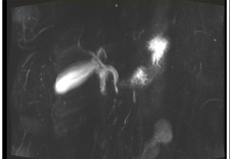
Lab results indicated hemoglobin at 12.8 gm/dl, leukocyte count at 7,050/mm3, platelets at 375,000/mm3, total bilirubin at 0.7 mg/dl, AST at 39 U/L, ALT at 54 U/L, and ALP at 155 U/L. MRCP revealed an extrahepatic biliary ductal structure, with two ducts converging in the lower part of the pancreatic head, typical of a type Va variant. Motion artifacts proved difficult to identify whether the anatomy represented a long cystic duct with a low insertion or a very low common bile duct bifurcation. The pancreatic lesion hadn't changed in size from images taken 2 years prior, not requiring medical intervention.
Discussion: We report a case of two long extrahepatic bile ducts joining distally to form one common bile duct with a long cystic duct joining the right extrahepatic duct at a low insertion. Embryogenesis of the hepatobiliary system occurs in the 3rd week of gestation. DCBD has been postulated to be a byproduct of multiple mechanisms, such as random subdivision of hepatic diverticulum, defect in the recanalization process, and disruption of development leading to the persistence of an extrahepatic accessory duct that later regresses to form a single common bile duct.

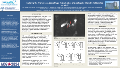
Figure: 1. MRCP showing two separate CBDs (right and left) running parallel and joining distally to form a single bile duct (type Va DCBD)
Disclosures:
Raj Mohan Ram Mohan indicated no relevant financial relationships.
Winghang Lau indicated no relevant financial relationships.
Sai Reshma Magam indicated no relevant financial relationships.
Sai Greeshma Magam indicated no relevant financial relationships.
Melvin Joy indicated no relevant financial relationships.
Dilman Natt indicated no relevant financial relationships.
Abhishek Tadikonda indicated no relevant financial relationships.
Leeza Pannikodu indicated no relevant financial relationships.
Jiten Desai indicated no relevant financial relationships.
Paul Mustacchia indicated no relevant financial relationships.
Raj Mohan Ram Mohan, MD, Winghang Lau, MD, Sai Reshma Magam, MD, Sai Greeshma Magam, MD, Melvin Joy, MD, Dilman Natt, MD, Abhishek Tadikonda, MD, Leeza Pannikodu, MD, Jiten Desai, MD, Paul Mustacchia, MD, MBA. P0102 - Exploring the Anomalies: A Case of Type Va Duplication of Extrahepatic Biliary Ducts Identified on MRCP, ACG 2024 Annual Scientific Meeting Abstracts. Philadelphia, PA: American College of Gastroenterology.
