Sunday Poster Session
Category: Biliary/Pancreas
P0148 - A Rare Case of Isolated Metastasis to the Pleura in the Setting of Newly Diagnosed Pancreatic Adenocarcinoma
Sunday, October 27, 2024
3:30 PM - 7:00 PM ET
Location: Exhibit Hall E

Has Audio

Rakahn Haddadin, MD
MountainView Hospital
Las Vegas, NV
Presenting Author(s)
Rakahn Haddadin, MD1, Humzah Iqbal, MD2, Danny Aboujamra, MD3, George Trad, MD4, John Ryan, MD4
1MountainView Hospital, Las Vegas, NV; 2University of California San Francisco, Fresno, CA; 3Medical University of South Carolina (MUSC) Health Florence, North York, ON, Canada; 4Sunrise Health GME Consortium, Las Vegas, NV
Introduction: Pancreatic cancer represents a formidable challenge in oncology due to its aggressive nature and often asymptomatic early stages. We present a patient with newly diagnosed pancreatic adenocarcinoma and new-onset isolated pleural metastasis. Pleural metastasis as an extra-pancreatic manifestation of pancreatic cancer is notably rare; its exact incidence is unknown.
Case Description/Methods: A 78-year-old female with a past medical history of gastroesophageal reflux disease (GERD), type 2 diabetes mellitus, chronic obstructive pulmonary disease (COPD), and recently diagnosed pancreatic adenocarcinoma presented to the ER due to worsening shortness of breath and cough. The patient had been diagnosed with pancreatic adenocarcinoma a few months prior to admission and had not started chemotherapy or radiation.
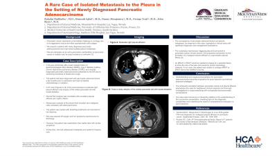
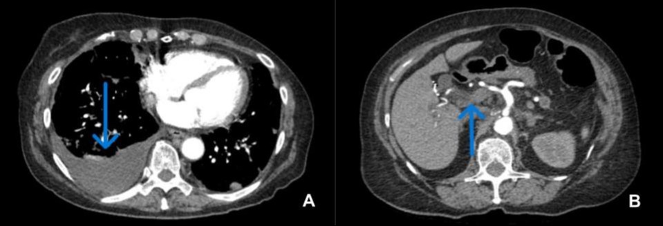
A CT scan (Figures A, B) of the chest showed a moderate right pleural effusion and atrophy of the visible pancreatic tail with ductal dilatation. Pleural fluid analysis was consistent with exudative pleural effusion per Light's criteria. Microscopic analysis of the pleural fluid revealed rare malignant cells consistent with adenocarcinoma.
The patient was treated with breathing treatments and steroids for her dyspnea. She was weaned off of oxygen and her symptoms resolved prior to discharge. However, the patient was readmitted a few weeks later with similar symptoms. At that time, she had widespread metastasis and opted for hospice care.
Discussion: The convergence of pancreatic adenocarcinoma and pleural metastasis, as observed in this case, represents a clinical rarity with significant diagnostic and management implications. The underlying mechanisms triggering pleural involvement in pancreatic cancer remain elusive. In this case, the patient initially presented with classic symptoms of COPD exacerbation and was found to have a malignant pleural effusion. One proposed mechanism for the formation of a malignant pleural effusion is a pancreatico-pleural fistula. A Magnetic resonance cholangiopancreatography (MRCP) or Endoscopic retrograde cholangiopancreatography (ERCP) would be needed to assess for a possible fistula to show the site of the leak and reveal the ductal morphology. However, in our case, the patient was unable to undergo MRCP or ERCP due to her inability to lay flat.

Disclosures:
Rakahn Haddadin, MD1, Humzah Iqbal, MD2, Danny Aboujamra, MD3, George Trad, MD4, John Ryan, MD4. P0148 - A Rare Case of Isolated Metastasis to the Pleura in the Setting of Newly Diagnosed Pancreatic Adenocarcinoma, ACG 2024 Annual Scientific Meeting Abstracts. Philadelphia, PA: American College of Gastroenterology.
1MountainView Hospital, Las Vegas, NV; 2University of California San Francisco, Fresno, CA; 3Medical University of South Carolina (MUSC) Health Florence, North York, ON, Canada; 4Sunrise Health GME Consortium, Las Vegas, NV
Introduction: Pancreatic cancer represents a formidable challenge in oncology due to its aggressive nature and often asymptomatic early stages. We present a patient with newly diagnosed pancreatic adenocarcinoma and new-onset isolated pleural metastasis. Pleural metastasis as an extra-pancreatic manifestation of pancreatic cancer is notably rare; its exact incidence is unknown.
Case Description/Methods: A 78-year-old female with a past medical history of gastroesophageal reflux disease (GERD), type 2 diabetes mellitus, chronic obstructive pulmonary disease (COPD), and recently diagnosed pancreatic adenocarcinoma presented to the ER due to worsening shortness of breath and cough. The patient had been diagnosed with pancreatic adenocarcinoma a few months prior to admission and had not started chemotherapy or radiation.
A CT scan (Figures A, B) of the chest showed a moderate right pleural effusion and atrophy of the visible pancreatic tail with ductal dilatation. Pleural fluid analysis was consistent with exudative pleural effusion per Light's criteria. Microscopic analysis of the pleural fluid revealed rare malignant cells consistent with adenocarcinoma.
The patient was treated with breathing treatments and steroids for her dyspnea. She was weaned off of oxygen and her symptoms resolved prior to discharge. However, the patient was readmitted a few weeks later with similar symptoms. At that time, she had widespread metastasis and opted for hospice care.
Discussion: The convergence of pancreatic adenocarcinoma and pleural metastasis, as observed in this case, represents a clinical rarity with significant diagnostic and management implications. The underlying mechanisms triggering pleural involvement in pancreatic cancer remain elusive. In this case, the patient initially presented with classic symptoms of COPD exacerbation and was found to have a malignant pleural effusion. One proposed mechanism for the formation of a malignant pleural effusion is a pancreatico-pleural fistula. A Magnetic resonance cholangiopancreatography (MRCP) or Endoscopic retrograde cholangiopancreatography (ERCP) would be needed to assess for a possible fistula to show the site of the leak and reveal the ductal morphology. However, in our case, the patient was unable to undergo MRCP or ERCP due to her inability to lay flat.

Figure: Figure A= Moderate right pleural effusion.
Figure B=Atrophy of the visible pancreatic tail with ductal dilatation.
Figure B=Atrophy of the visible pancreatic tail with ductal dilatation.
Disclosures:
Rakahn Haddadin indicated no relevant financial relationships.
Humzah Iqbal indicated no relevant financial relationships.
Danny Aboujamra indicated no relevant financial relationships.
George Trad indicated no relevant financial relationships.
John Ryan indicated no relevant financial relationships.
Rakahn Haddadin, MD1, Humzah Iqbal, MD2, Danny Aboujamra, MD3, George Trad, MD4, John Ryan, MD4. P0148 - A Rare Case of Isolated Metastasis to the Pleura in the Setting of Newly Diagnosed Pancreatic Adenocarcinoma, ACG 2024 Annual Scientific Meeting Abstracts. Philadelphia, PA: American College of Gastroenterology.
