Sunday Poster Session
Category: Colon
P0213 - Incidental Colon Adenoma Detection on FDG PET-CT
Sunday, October 27, 2024
3:30 PM - 7:00 PM ET
Location: Exhibit Hall E


Anoushka Dua, MD
UC Irvine Health
Orange, CA
Presenting Author(s)
Anoushka Dua, MD1, William Karnes, MD1, Valery Vilchez Parra, MD2
1UC Irvine Health, Orange, CA; 2UCI, Orange, CA
Introduction: It is well known that abnormal FDG PET-CT imaging of the colon frequently prompts colonoscopy, resulting in the incidental detection of adenomas. Few studies in the last decade have reported on the sensitivity of FDG PET-CT in the detection of colon adenomas, despite advancements in this imaging modality in recent years.
Methods: We conducted a retrospective chart review of patients who underwent colonoscopy with the indication of “abnormal PET” between 9/27/2012 and 3/1/2024 using an initial query within our institution’s colonoscopy quality database. Retrospective chart review for each patient confirmed age, sex, and procedure indication of abnormal PET. Cases with abnormal PET of the GI tract outside of the colon were excluded. We abstracted the most recent PET findings, colonoscopy findings, and pathology findings. Each case was characterized as one of the following: endoscopic polyp correlate for PET findings, endoscopic non-polyp correlate for PET findings, or no endoscopic correlate for PET findings. We utilized descriptive statistics to calculate the frequency of each category, characterize and quantify the polyps detected and not detected by PET, and calculate the adenoma, advanced adenoma, and cancer detection rate.
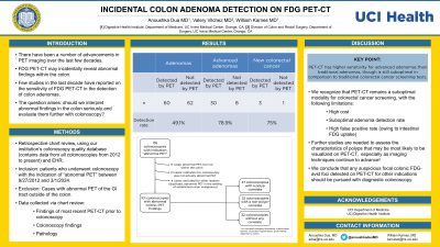
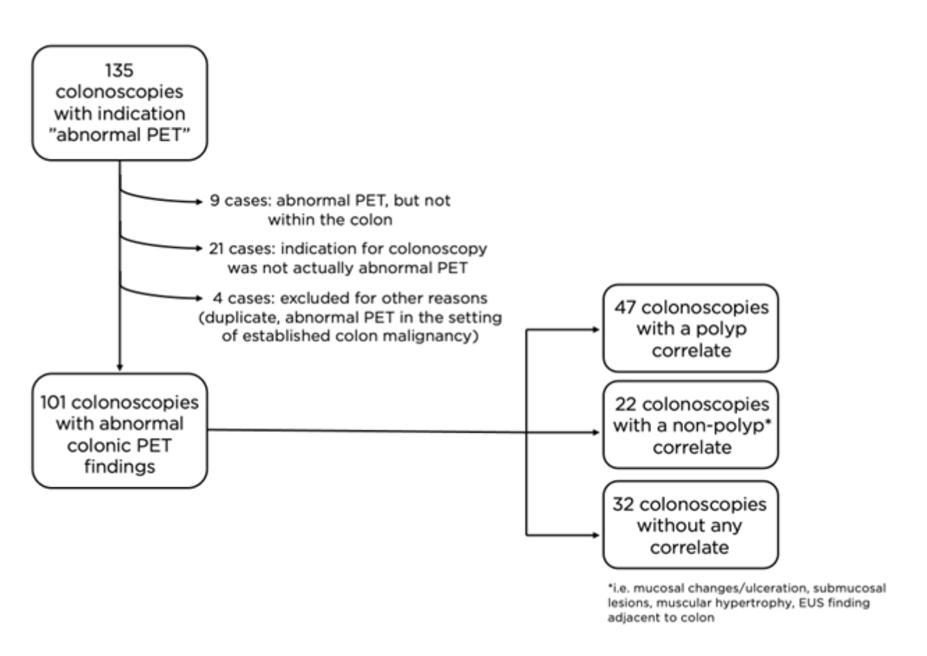
Results: 135 cases were generated from the initial query. Of these, manual chart review excluded 34 cases that did not meet inclusion criteria (Figure). There was an endoscopic polyp correlate for abnormal PET findings in 47 cases (46.5%), with a total of 64 polyps detected by PET, 30 of which were advanced adenomas, as well as 3 cases of new colorectal cancer. There was an endoscopic non-polyp correlate in 22 cases (21.8%), and no endoscopic correlate in 32 cases (31.7%). There were 68 polyps not detected by PET, of which 8 were advanced adenomas (Table). The overall adenoma detection rate on PET was 49.1%, and the advanced adenoma detection rate was 79%. There was one cancerous lesion (a case of intramucosal adenocarcinoma arising from TVA with negative margins) not detected by PET.
Discussion: We recognize that PET-CT remains a suboptimal modality for colorectal cancer screening owing to its high cost and suboptimal adenoma detection rate. Further studies are needed to assess the characteristics of polyps that may be most likely to be visualized on PET-CT, especially as imaging techniques and quality continue to be advanced. Regardless, any focal colonic FDG-avid foci detected on PET-CT for other indications should be pursued with diagnostic colonoscopy.

Note: The table for this abstract can be viewed in the ePoster Gallery section of the ACG 2024 ePoster Site or in The American Journal of Gastroenterology's abstract supplement issue, both of which will be available starting October 27, 2024.
Disclosures:
Anoushka Dua, MD1, William Karnes, MD1, Valery Vilchez Parra, MD2. P0213 - Incidental Colon Adenoma Detection on FDG PET-CT, ACG 2024 Annual Scientific Meeting Abstracts. Philadelphia, PA: American College of Gastroenterology.
1UC Irvine Health, Orange, CA; 2UCI, Orange, CA
Introduction: It is well known that abnormal FDG PET-CT imaging of the colon frequently prompts colonoscopy, resulting in the incidental detection of adenomas. Few studies in the last decade have reported on the sensitivity of FDG PET-CT in the detection of colon adenomas, despite advancements in this imaging modality in recent years.
Methods: We conducted a retrospective chart review of patients who underwent colonoscopy with the indication of “abnormal PET” between 9/27/2012 and 3/1/2024 using an initial query within our institution’s colonoscopy quality database. Retrospective chart review for each patient confirmed age, sex, and procedure indication of abnormal PET. Cases with abnormal PET of the GI tract outside of the colon were excluded. We abstracted the most recent PET findings, colonoscopy findings, and pathology findings. Each case was characterized as one of the following: endoscopic polyp correlate for PET findings, endoscopic non-polyp correlate for PET findings, or no endoscopic correlate for PET findings. We utilized descriptive statistics to calculate the frequency of each category, characterize and quantify the polyps detected and not detected by PET, and calculate the adenoma, advanced adenoma, and cancer detection rate.
Results: 135 cases were generated from the initial query. Of these, manual chart review excluded 34 cases that did not meet inclusion criteria (Figure). There was an endoscopic polyp correlate for abnormal PET findings in 47 cases (46.5%), with a total of 64 polyps detected by PET, 30 of which were advanced adenomas, as well as 3 cases of new colorectal cancer. There was an endoscopic non-polyp correlate in 22 cases (21.8%), and no endoscopic correlate in 32 cases (31.7%). There were 68 polyps not detected by PET, of which 8 were advanced adenomas (Table). The overall adenoma detection rate on PET was 49.1%, and the advanced adenoma detection rate was 79%. There was one cancerous lesion (a case of intramucosal adenocarcinoma arising from TVA with negative margins) not detected by PET.
Discussion: We recognize that PET-CT remains a suboptimal modality for colorectal cancer screening owing to its high cost and suboptimal adenoma detection rate. Further studies are needed to assess the characteristics of polyps that may be most likely to be visualized on PET-CT, especially as imaging techniques and quality continue to be advanced. Regardless, any focal colonic FDG-avid foci detected on PET-CT for other indications should be pursued with diagnostic colonoscopy.

Figure: Summary of colonoscopies performed for indication of abnormal PET-CT of the colon, and their endoscopic findings.
Note: The table for this abstract can be viewed in the ePoster Gallery section of the ACG 2024 ePoster Site or in The American Journal of Gastroenterology's abstract supplement issue, both of which will be available starting October 27, 2024.
Disclosures:
Anoushka Dua indicated no relevant financial relationships.
William Karnes indicated no relevant financial relationships.
Valery Vilchez Parra indicated no relevant financial relationships.
Anoushka Dua, MD1, William Karnes, MD1, Valery Vilchez Parra, MD2. P0213 - Incidental Colon Adenoma Detection on FDG PET-CT, ACG 2024 Annual Scientific Meeting Abstracts. Philadelphia, PA: American College of Gastroenterology.
