Sunday Poster Session
Category: Colon
P0254 - What Can Cecal Nodules Be?
Sunday, October 27, 2024
3:30 PM - 7:00 PM ET
Location: Exhibit Hall E

Has Audio

Busara Songtanin, MD
Texas Tech University Health Sciences Center
Lubbock, TX
Presenting Author(s)
Busara Songtanin, MD1, Austin Miller, MD2, Daoud Arif, MD1, Vanessa Costilla, MD2
1Texas Tech University Health Sciences Center, Lubbock, TX; 2University Medical Center, Lubbock, TX
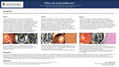
Introduction: Subepithelial lesions of the gastrointestinal tract can be benign or malignant. Managing these lesions can be challenging. We present a case series of three patients with subepithelial nodules in the cecum discovered during colonoscopy.
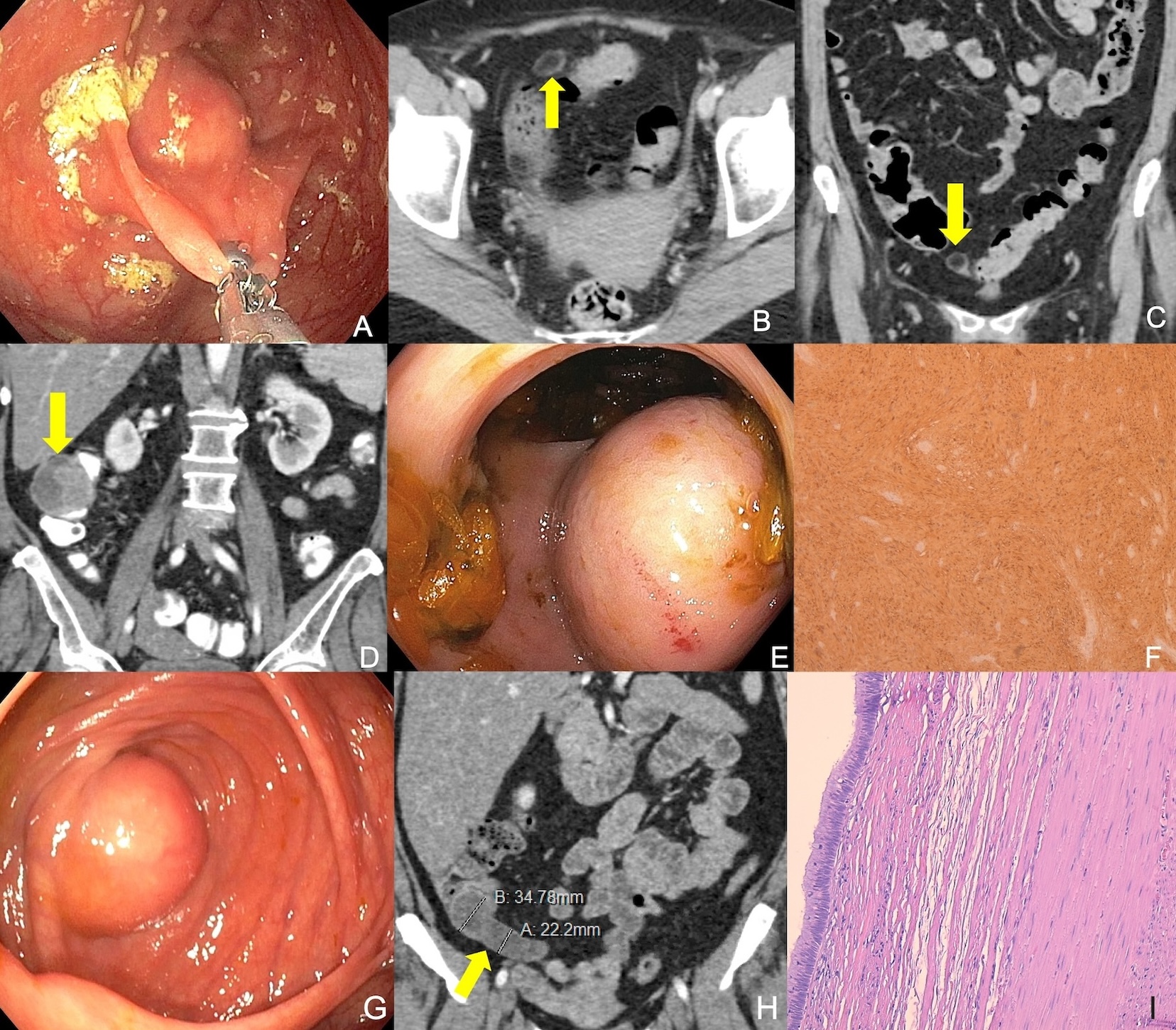
Case Description/Methods: Case 1: A 42-year-old female with a history of breast cancer underwent colonoscopy for surveillance. The colonoscopy revealed a 9mm subepithelial nodule in the cecum (Figure A). Pathology showed benign colonic mucosa. Computed tomography (CT) of the abdomen and pelvis with contrast following colonoscopy showed inflammatory changes surrounding the sigmoid colon suggesting epiploic appendagitis (Figure B and C). Surgical consultation led to combined endoscopic-laparoscopic surgery, revealing a hardened epiploic appendage compressing against the wall of the cecum. The patient recovered well at one month follow-up.
Case 2: An 82-year-old female with a history of anemia of chronic disease underwent a CT abdomen and pelvis with contrast workup for anemia. The CT showed a 5 cm x 4.5 cm exophytic heterogeneous mass arising from the cecum (Figure D). A colonoscopy revealed a 4cm x 5cm subepithelial mass in the cecum with no bleeding (Figure E). Pathology showed benign colonic mucosa. Surgery was consulted and the patient underwent diagnostic laparoscopy for a right colectomy. Pathology of the mass showed schwannoma (Figure 1F), and the patient recovered well at two weeks post-surgery.
Case 3: A 55-year-old woman with a history of hypertension underwent colonoscopy surveillance. A colonoscopy showed a 3cm subepithelial nodule at the appendiceal orifice (Figure G). Pathology showed benign colonic mucosa. The CT abdomen with contrast showed a 3 cm cystic structure in the cecum (Figure H). Surgery was consulted, and the patient underwent diagnostic laparoscopy for a right colectomy. Pathology showed a low-grade appendiceal mucinous neoplasm (Figure I). The patient recovered well at one month follow-up.
Discussion: The challenge in managing subepithelial lesions in the intestine is that the tissue obtained during standard colonoscopy can be non-diagnostic. The current guideline recommends using endoscopic ultrasound with tissue acquisition to help determine tissue characteristics. Other techniques, such as combined endoscopic-laparoscopic surgery, have been proposed as an alternative to bowel resection in patients with large benign polyp(s) in a difficult anatomical location where endoscopic resection is not feasible.

Disclosures:
Busara Songtanin, MD1, Austin Miller, MD2, Daoud Arif, MD1, Vanessa Costilla, MD2. P0254 - What Can Cecal Nodules Be?, ACG 2024 Annual Scientific Meeting Abstracts. Philadelphia, PA: American College of Gastroenterology.
1Texas Tech University Health Sciences Center, Lubbock, TX; 2University Medical Center, Lubbock, TX
Introduction: Subepithelial lesions of the gastrointestinal tract can be benign or malignant. Managing these lesions can be challenging. We present a case series of three patients with subepithelial nodules in the cecum discovered during colonoscopy.
Case Description/Methods: Case 1: A 42-year-old female with a history of breast cancer underwent colonoscopy for surveillance. The colonoscopy revealed a 9mm subepithelial nodule in the cecum (Figure A). Pathology showed benign colonic mucosa. Computed tomography (CT) of the abdomen and pelvis with contrast following colonoscopy showed inflammatory changes surrounding the sigmoid colon suggesting epiploic appendagitis (Figure B and C). Surgical consultation led to combined endoscopic-laparoscopic surgery, revealing a hardened epiploic appendage compressing against the wall of the cecum. The patient recovered well at one month follow-up.
Case 2: An 82-year-old female with a history of anemia of chronic disease underwent a CT abdomen and pelvis with contrast workup for anemia. The CT showed a 5 cm x 4.5 cm exophytic heterogeneous mass arising from the cecum (Figure D). A colonoscopy revealed a 4cm x 5cm subepithelial mass in the cecum with no bleeding (Figure E). Pathology showed benign colonic mucosa. Surgery was consulted and the patient underwent diagnostic laparoscopy for a right colectomy. Pathology of the mass showed schwannoma (Figure 1F), and the patient recovered well at two weeks post-surgery.
Case 3: A 55-year-old woman with a history of hypertension underwent colonoscopy surveillance. A colonoscopy showed a 3cm subepithelial nodule at the appendiceal orifice (Figure G). Pathology showed benign colonic mucosa. The CT abdomen with contrast showed a 3 cm cystic structure in the cecum (Figure H). Surgery was consulted, and the patient underwent diagnostic laparoscopy for a right colectomy. Pathology showed a low-grade appendiceal mucinous neoplasm (Figure I). The patient recovered well at one month follow-up.
Discussion: The challenge in managing subepithelial lesions in the intestine is that the tissue obtained during standard colonoscopy can be non-diagnostic. The current guideline recommends using endoscopic ultrasound with tissue acquisition to help determine tissue characteristics. Other techniques, such as combined endoscopic-laparoscopic surgery, have been proposed as an alternative to bowel resection in patients with large benign polyp(s) in a difficult anatomical location where endoscopic resection is not feasible.

Figure: (A): Colonoscopy shows a 9 mm subepithelial nodule at the cecum.
(B) and (C): CT abdomen and pelvis with contrast axial plane and coronal plane shows inflammatory changes surrounding the sigmoid colon which suggest an epiploic appendagitis (yellow arrow).
(D): CT abdomen and pelvis with contrast coronal plane shows a 4.5 cm x 5 cm exophytic heterogenous mass with cystic contest arising from the ascending colon (yellow arrow).
(E): Colonoscopy shows a 4 cm x 5 cm subepithelial non-obstructing large mass at the cecum with no bleeding or ulcer. Negative pillow sign.
(F): Histopathology stain of the biopsy shows diffusely and strongly positive S100 stain.
(G): Colonoscopy shows a 3cm subepithelial nodule at the cecum.
(H): CT abdomen and pelvis with contrast shows a fluid density mass measuring 3cm near the appendiceal orifice with dilatation of the appendix measuring 2.1cm in diameter (yellow arrow). There is no significant peri-appendiceal inflammatory changes.
(I): Hematoxylin and Eosin stain shows cells with abundant apical mucin, elongated nuclei, loss of lamina propria and lymphoid aggregates, consistent with low-grade appendiceal mucinous neoplasm
Disclosures:
Busara Songtanin indicated no relevant financial relationships.
Austin Miller indicated no relevant financial relationships.
Daoud Arif indicated no relevant financial relationships.
Vanessa Costilla indicated no relevant financial relationships.
Busara Songtanin, MD1, Austin Miller, MD2, Daoud Arif, MD1, Vanessa Costilla, MD2. P0254 - What Can Cecal Nodules Be?, ACG 2024 Annual Scientific Meeting Abstracts. Philadelphia, PA: American College of Gastroenterology.
