Sunday Poster Session
Category: Colon
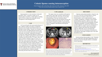
P0256 - Colonic Lipoma Causing Intussusception
Sunday, October 27, 2024
3:30 PM - 7:00 PM ET
Location: Exhibit Hall E

Has Audio

Busara Songtanin, MD
Texas Tech University Health Sciences Center
Lubbock, TX
Presenting Author(s)
Busara Songtanin, MD1, Afrina Rimu, MD1, Dauod Arid, MD1, Vanessa Costilla, MD2
1Texas Tech University Health Sciences Center, Lubbock, TX; 2University Medical Center, Lubbock, TX
Introduction: Intussusception is a rare occurrence in the adult population. Here, we present a unique case of intussusception caused by a colonic lipoma, shedding light on the less common but potentially serious consequences of this benign growth.
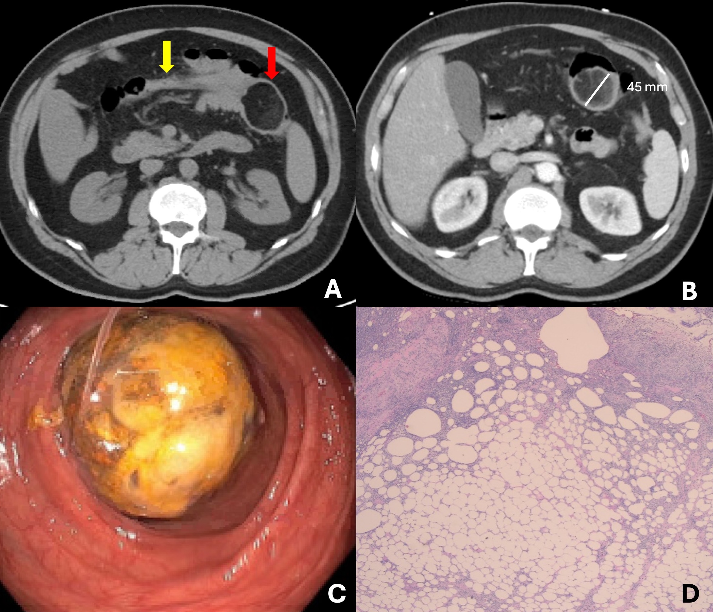
Case Description/Methods: A 62-year-old male with a past medical history of type 2 diabetes mellitus and chronic constipation presented to the hospital with abdominal pain and bloating for three months. The pain was constant. Over the past two days, the abdominal pain became more severe and was associated with nausea and vomiting. Vital signs were normal. Abdominal examination revealed soft, mildly distended abdomen, tenderness at the right lower quadrant, and hypoactive bowel sounds. Laboratory tests showed Hb of 15.0 g/dL, white cell count of 6.9k/µL (reference range 4-11 k/µL). An abdominal X-ray revealed moderate stool in the colon with a non-obstructing bowel gas pattern. CT abdomen with and without intravenous contrast showed a 3.5cm x 4.5 cm fat density structure within the transverse colon, suspected to be a low-grade lipoma resulting in an intussusception (Figure A and B). Colonoscopy showed a polypoid, non-circumferential, ulcerated mass measuring 50mm in diameter, partially obstructing the distal transverse colon (Figure C). The surgery team was consulted, and the patient underwent an open right colectomy, during which a 4cm proximal transverse colon mass was revealed. Histopathology of the mass indicated a bland lipomatous neoplasm with associated fat necrosis and surrounding inflammation (Figure D). Two months post-surgery, the patient reported no abdominal pain, and his constipation and abdominal bloating had improved.
Discussion: A lipoma causing intussusception is found in only 0.5-4.4% of all cases of intussusception. Clinical presentations are similar to other causes of intussusception, such as abdominal pain, nausea, vomiting, constipation, or changes in bowel habits. Intussusception caused by a lipoma can be easily diagnosed with abdominal ultrasonography, although computed tomography is the gold standard for an accurate diagnosis. In contrast to the management of intussusception in children, adult intussusception requires a surgical and endoscopic management approach. Minimally invasive techniques like endoscopic removal of the lipoma are preferred for reducing pain and length of hospital stay in the small lipomatous lesions.

Disclosures:
Busara Songtanin, MD1, Afrina Rimu, MD1, Dauod Arid, MD1, Vanessa Costilla, MD2. P0256 - Colonic Lipoma Causing Intussusception, ACG 2024 Annual Scientific Meeting Abstracts. Philadelphia, PA: American College of Gastroenterology.
1Texas Tech University Health Sciences Center, Lubbock, TX; 2University Medical Center, Lubbock, TX
Introduction: Intussusception is a rare occurrence in the adult population. Here, we present a unique case of intussusception caused by a colonic lipoma, shedding light on the less common but potentially serious consequences of this benign growth.
Case Description/Methods: A 62-year-old male with a past medical history of type 2 diabetes mellitus and chronic constipation presented to the hospital with abdominal pain and bloating for three months. The pain was constant. Over the past two days, the abdominal pain became more severe and was associated with nausea and vomiting. Vital signs were normal. Abdominal examination revealed soft, mildly distended abdomen, tenderness at the right lower quadrant, and hypoactive bowel sounds. Laboratory tests showed Hb of 15.0 g/dL, white cell count of 6.9k/µL (reference range 4-11 k/µL). An abdominal X-ray revealed moderate stool in the colon with a non-obstructing bowel gas pattern. CT abdomen with and without intravenous contrast showed a 3.5cm x 4.5 cm fat density structure within the transverse colon, suspected to be a low-grade lipoma resulting in an intussusception (Figure A and B). Colonoscopy showed a polypoid, non-circumferential, ulcerated mass measuring 50mm in diameter, partially obstructing the distal transverse colon (Figure C). The surgery team was consulted, and the patient underwent an open right colectomy, during which a 4cm proximal transverse colon mass was revealed. Histopathology of the mass indicated a bland lipomatous neoplasm with associated fat necrosis and surrounding inflammation (Figure D). Two months post-surgery, the patient reported no abdominal pain, and his constipation and abdominal bloating had improved.
Discussion: A lipoma causing intussusception is found in only 0.5-4.4% of all cases of intussusception. Clinical presentations are similar to other causes of intussusception, such as abdominal pain, nausea, vomiting, constipation, or changes in bowel habits. Intussusception caused by a lipoma can be easily diagnosed with abdominal ultrasonography, although computed tomography is the gold standard for an accurate diagnosis. In contrast to the management of intussusception in children, adult intussusception requires a surgical and endoscopic management approach. Minimally invasive techniques like endoscopic removal of the lipoma are preferred for reducing pain and length of hospital stay in the small lipomatous lesions.

Figure: Figure 1
(A) Axial section of CT abdomen without contrast an intussusception of transverse colon (yellow arrow) into a lipoma (red arrow).
(B) Axial section of CT abdomen with intravenous contrast showed a 3.5x4.5cm fat density structure within the transverse colon (yellow arrow).
(C) Colonoscopy showed a non-circumferential obstructing mass measured 5cm at the distal transverse colon.
(D) Histology shows a bland lipomatous neoplasm consisting of mature benign adipose tissue effacing mucosa and submucosa of the intestine with associated fat necrosis and surrounding inflammation.
Disclosures:
Busara Songtanin indicated no relevant financial relationships.
Afrina Rimu indicated no relevant financial relationships.
Dauod Arid indicated no relevant financial relationships.
Vanessa Costilla indicated no relevant financial relationships.
Busara Songtanin, MD1, Afrina Rimu, MD1, Dauod Arid, MD1, Vanessa Costilla, MD2. P0256 - Colonic Lipoma Causing Intussusception, ACG 2024 Annual Scientific Meeting Abstracts. Philadelphia, PA: American College of Gastroenterology.
