Sunday Poster Session
Category: Colon
P0278 - Colon Adenocarcinoma Presenting With Peri-Colonic Abscess Formation
Sunday, October 27, 2024
3:30 PM - 7:00 PM ET
Location: Exhibit Hall E

Has Audio

Dakota Bigham, MD
University of Texas Health San Antonio
San Antonio, TX
Presenting Author(s)
Dakota Bigham, MD1, Abakar Baraka, 2, Elizabeth Bullo, 2, Jonathan Selzman, DO2, Jawairia Memon, MD3
1University of Texas Health San Antonio, San Antonio, TX; 2UTHSCSA, San Antonio, TX; 3University of Texas Health Science Center, San Antonio, TX
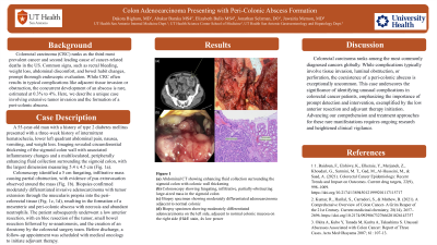
Introduction: Colorectal carcinoma (CRC) ranks as the third most prevalent cancer and second leading cause of cancer-related deaths in the US. Common signs, such as rectal bleeding, weight loss, abdominal discomfort, and bowel habit changes, prompt thorough endoscopic evaluation. While CRC often results in typical complications like adjacent tissue invasion or obstruction, the concurrent development of an abscess is rare, estimated at 0.3% to 4%. Here, we describe a unique case involving extensive tumor invasion and the formation of a peri-colonic abscess.
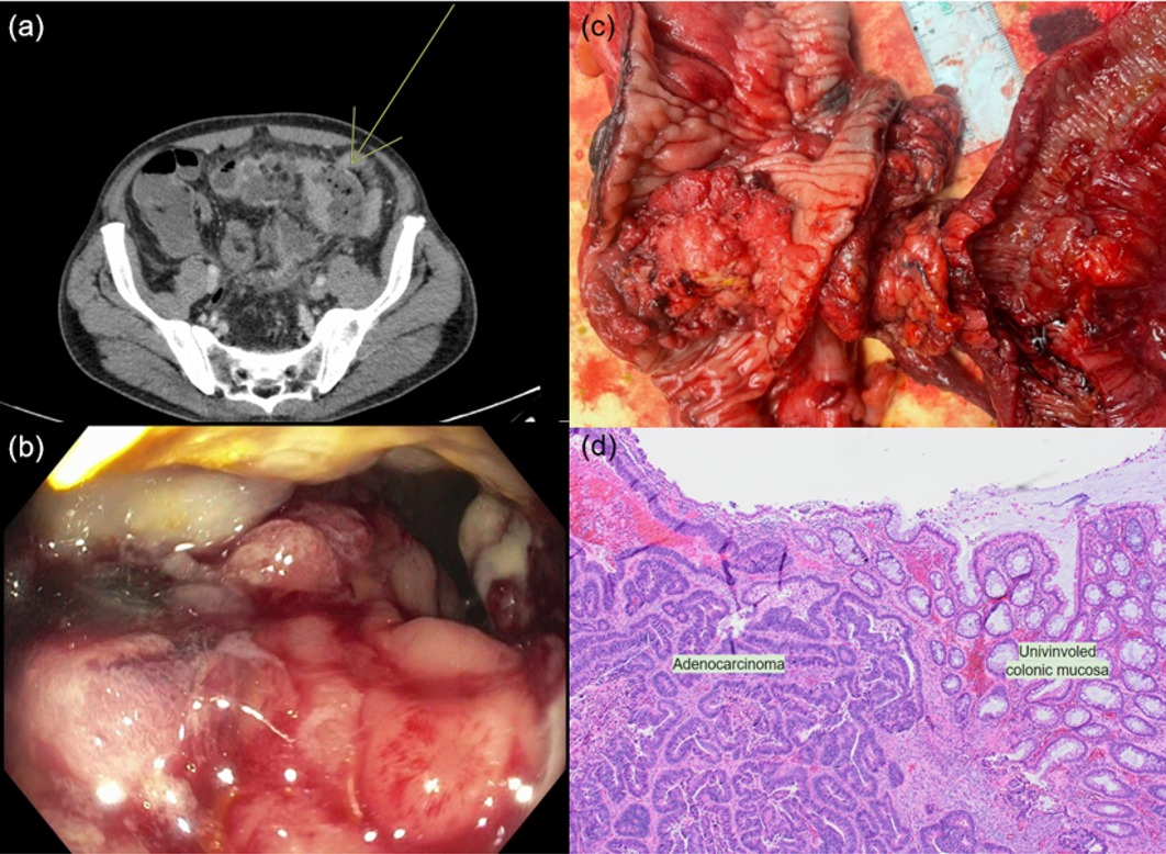
Case Description/Methods: A 55-year-old man with a history of type 2 diabetes mellitus presented with a three-week history of intermittent hematochezia, lower left quadrant abdominal pain, nausea, vomiting, and weight loss. Imaging revealed circumferential thickening of the sigmoid colon wall with associated inflammatory changes and a multiloculated, peripherally enhancing fluid collection surrounding the sigmoid colon, with the largest dimension measuring 5.4 x 4.5 cm (Fig. 1a).
Colonoscopy identified a 5 cm fungating, infiltrative mass causing partial obstruction, with evidence of pus extravasation observed around the mass (Fig. 1b). Biopsies confirmed moderately differentiated invasive adenocarcinoma with tumor extension through the muscularis propria into the peri-colorectal tissue (Fig. 1c, 1d), resulting in the formation of a mesenteric and peri-colonic abscess with necrosis and abundant neutrophils.
The patient subsequently underwent a low anterior resection, with en bloc resection of the tumor, small bowel resection followed by re-anastomosis, and the creation of an ileostomy by the colorectal surgery team. Before discharge, a follow-up appointment was scheduled with medical oncology to initiate adjuvant therapy.
Discussion: Colorectal carcinoma ranks among the most commonly diagnosed cancers globally. While complications typically involve tissue invasion, luminal obstruction, or perforation, the coexistence of a peri-colonic abscess is exceptionally uncommon. This case underscores the significance of identifying unusual complications in colorectal cancer patients, emphasizing the importance of prompt detection and intervention, exemplified by the low anterior resection and adjuvant therapy initiation. Advancing our comprehension and treatment approaches for these rare manifestations requires ongoing research and heightened clinical vigilance.

Disclosures:
Dakota Bigham, MD1, Abakar Baraka, 2, Elizabeth Bullo, 2, Jonathan Selzman, DO2, Jawairia Memon, MD3. P0278 - Colon Adenocarcinoma Presenting With Peri-Colonic Abscess Formation, ACG 2024 Annual Scientific Meeting Abstracts. Philadelphia, PA: American College of Gastroenterology.
1University of Texas Health San Antonio, San Antonio, TX; 2UTHSCSA, San Antonio, TX; 3University of Texas Health Science Center, San Antonio, TX
Introduction: Colorectal carcinoma (CRC) ranks as the third most prevalent cancer and second leading cause of cancer-related deaths in the US. Common signs, such as rectal bleeding, weight loss, abdominal discomfort, and bowel habit changes, prompt thorough endoscopic evaluation. While CRC often results in typical complications like adjacent tissue invasion or obstruction, the concurrent development of an abscess is rare, estimated at 0.3% to 4%. Here, we describe a unique case involving extensive tumor invasion and the formation of a peri-colonic abscess.
Case Description/Methods: A 55-year-old man with a history of type 2 diabetes mellitus presented with a three-week history of intermittent hematochezia, lower left quadrant abdominal pain, nausea, vomiting, and weight loss. Imaging revealed circumferential thickening of the sigmoid colon wall with associated inflammatory changes and a multiloculated, peripherally enhancing fluid collection surrounding the sigmoid colon, with the largest dimension measuring 5.4 x 4.5 cm (Fig. 1a).
Colonoscopy identified a 5 cm fungating, infiltrative mass causing partial obstruction, with evidence of pus extravasation observed around the mass (Fig. 1b). Biopsies confirmed moderately differentiated invasive adenocarcinoma with tumor extension through the muscularis propria into the peri-colorectal tissue (Fig. 1c, 1d), resulting in the formation of a mesenteric and peri-colonic abscess with necrosis and abundant neutrophils.
The patient subsequently underwent a low anterior resection, with en bloc resection of the tumor, small bowel resection followed by re-anastomosis, and the creation of an ileostomy by the colorectal surgery team. Before discharge, a follow-up appointment was scheduled with medical oncology to initiate adjuvant therapy.
Discussion: Colorectal carcinoma ranks among the most commonly diagnosed cancers globally. While complications typically involve tissue invasion, luminal obstruction, or perforation, the coexistence of a peri-colonic abscess is exceptionally uncommon. This case underscores the significance of identifying unusual complications in colorectal cancer patients, emphasizing the importance of prompt detection and intervention, exemplified by the low anterior resection and adjuvant therapy initiation. Advancing our comprehension and treatment approaches for these rare manifestations requires ongoing research and heightened clinical vigilance.

Figure: Figure 1: a) Abdominal enhanced CT showing enhancing fluid collection surrounding the sigmoid colon with colonic wall thickening. b) Colonoscopy showing fungating, infiltrative, partially-obstructive mass in the sigmoid colon. c) Biopsy specimen showing moderately differentiated adenocarcinoma adjacent to normal colon. d) Biopsy specimen showing moderately differentiated adenocarcinoma on the left side, adjacent to normal colonic mucosa on the right side (H&E stain, 4x low power)
Disclosures:
Dakota Bigham indicated no relevant financial relationships.
Abakar Baraka indicated no relevant financial relationships.
Elizabeth Bullo indicated no relevant financial relationships.
Jonathan Selzman indicated no relevant financial relationships.
Jawairia Memon indicated no relevant financial relationships.
Dakota Bigham, MD1, Abakar Baraka, 2, Elizabeth Bullo, 2, Jonathan Selzman, DO2, Jawairia Memon, MD3. P0278 - Colon Adenocarcinoma Presenting With Peri-Colonic Abscess Formation, ACG 2024 Annual Scientific Meeting Abstracts. Philadelphia, PA: American College of Gastroenterology.
