Sunday Poster Session
Category: Colon
P0322 - Pancreatic Metastasis from Primary Colon Adenocarcinoma Presenting as a Newly Diagnosed Type 2 Diabetic with Obstructive Symptoms
Sunday, October 27, 2024
3:30 PM - 7:00 PM ET
Location: Exhibit Hall E

Has Audio

Bridgette B. McNally, DO
Mayo Clinic School of Graduate Medical Education
Scottsdale, AZ
Presenting Author(s)
Bridgette B. McNally, DO1, Jewel Samadder, MD2, Danial Haris. Shaikh, MD3, Dora Lam-Himlin, MD4, Alessandra Ceolin Schmitt, MD4, Kumaresan Sandrasegaran, MBChB5, Rahul Pannala, MD4
1Mayo Clinic School of Graduate Medical Education, Scottsdale, AZ; 2Mayo Clinic College of Medicine and Science, Scottsdale, AZ; 3Mayo Clinic, Cave Creek, AZ; 4Mayo Clinic, Scottsdale, AZ; 5Mayo Clinic, Phoenix, AZ
Introduction: Only 20% of colon cancer cases present with metastases at time of diagnoses, commonly to the peritoneum, liver or lung. 94% of pancreatic cancers are primary in origin. We present a patient with new onset diabetes & bowel obstruction found to have pancreatic metastasis from primary colon adenocarcinoma.
Case Description/Methods: A 62-year-old male was found to have a blood glucose >300 without a history of diabetes, in the setting of 20 lb weight loss, abdominal pain & constipation. A CT abdomen & pelvis showed lung & adrenal masses, & focal circumferential wall thickening of the sigmoid colon. A colonoscopy was planned, but he noted vomiting when completing his bowel preparation, prompting presentation to our ER. On arrival, labs were notable for WBC 17.2, blood glucose 334. CEA was mildly elevated to 3.4 and CA 19-9 was normal. A CT abdomen and pelvis noted a non-constricting sigmoid colon mass with upstream obstruction, 1.6 cm pancreatic body mass with upstream dilation, and adrenal & lung lesions. Given the multiple lesions and extensive family history of malignancy, there was a concern for a hereditary cancer syndrome. Lung biopsy was concerning for metastatic adenocarcinoma. A flexible sigmoidoscopy revealed a fungating & obstructive mass 20 cm from the anal verge and a Wallstent was deployed. Colonic biopsy showed invasive adenocarcinoma. An endoscopic ultrasound showed a pancreatic body mass with biopsy of metastatic adenocarcinoma, favoring colonic primary. The patient was started on FOLFOXIRI for metastatic colon cancer.
Discussion: Only < 6% of pancreatic masses are metastases and most from a breast or renal cell primary. And, only 6% are from primary colon cancer and it hypothesized that metastasis occurs via lymphagogues spread. The metastatic lesions are typically hypodense on CT imaging, which is hard to distinguish from primary pancreatic lesions. Presentation varies with most cases being asymptomatic, but overall more symptomatic than patients with primary pancreatic cancer. One case has reported diagnosis of pancreatic metastasis from colonic primary occurring in the setting of new onset diabetes, with ketoacidosis. Management of these metastatic pancreatic lesions can be surgical with systemic chemotherapy or chemotherapy alone; however, there is limited data comparing these outcomes, given low incidence. In our case, careful analysis & tissue sampling identified a primary colon adenocarcinoma with multiple metastatic lesions, ensuring targeted therapy.

Disclosures:
Bridgette B. McNally, DO1, Jewel Samadder, MD2, Danial Haris. Shaikh, MD3, Dora Lam-Himlin, MD4, Alessandra Ceolin Schmitt, MD4, Kumaresan Sandrasegaran, MBChB5, Rahul Pannala, MD4. P0322 - Pancreatic Metastasis from Primary Colon Adenocarcinoma Presenting as a Newly Diagnosed Type 2 Diabetic with Obstructive Symptoms, ACG 2024 Annual Scientific Meeting Abstracts. Philadelphia, PA: American College of Gastroenterology.
1Mayo Clinic School of Graduate Medical Education, Scottsdale, AZ; 2Mayo Clinic College of Medicine and Science, Scottsdale, AZ; 3Mayo Clinic, Cave Creek, AZ; 4Mayo Clinic, Scottsdale, AZ; 5Mayo Clinic, Phoenix, AZ
Introduction: Only 20% of colon cancer cases present with metastases at time of diagnoses, commonly to the peritoneum, liver or lung. 94% of pancreatic cancers are primary in origin. We present a patient with new onset diabetes & bowel obstruction found to have pancreatic metastasis from primary colon adenocarcinoma.
Case Description/Methods: A 62-year-old male was found to have a blood glucose >300 without a history of diabetes, in the setting of 20 lb weight loss, abdominal pain & constipation. A CT abdomen & pelvis showed lung & adrenal masses, & focal circumferential wall thickening of the sigmoid colon. A colonoscopy was planned, but he noted vomiting when completing his bowel preparation, prompting presentation to our ER. On arrival, labs were notable for WBC 17.2, blood glucose 334. CEA was mildly elevated to 3.4 and CA 19-9 was normal. A CT abdomen and pelvis noted a non-constricting sigmoid colon mass with upstream obstruction, 1.6 cm pancreatic body mass with upstream dilation, and adrenal & lung lesions. Given the multiple lesions and extensive family history of malignancy, there was a concern for a hereditary cancer syndrome. Lung biopsy was concerning for metastatic adenocarcinoma. A flexible sigmoidoscopy revealed a fungating & obstructive mass 20 cm from the anal verge and a Wallstent was deployed. Colonic biopsy showed invasive adenocarcinoma. An endoscopic ultrasound showed a pancreatic body mass with biopsy of metastatic adenocarcinoma, favoring colonic primary. The patient was started on FOLFOXIRI for metastatic colon cancer.
Discussion: Only < 6% of pancreatic masses are metastases and most from a breast or renal cell primary. And, only 6% are from primary colon cancer and it hypothesized that metastasis occurs via lymphagogues spread. The metastatic lesions are typically hypodense on CT imaging, which is hard to distinguish from primary pancreatic lesions. Presentation varies with most cases being asymptomatic, but overall more symptomatic than patients with primary pancreatic cancer. One case has reported diagnosis of pancreatic metastasis from colonic primary occurring in the setting of new onset diabetes, with ketoacidosis. Management of these metastatic pancreatic lesions can be surgical with systemic chemotherapy or chemotherapy alone; however, there is limited data comparing these outcomes, given low incidence. In our case, careful analysis & tissue sampling identified a primary colon adenocarcinoma with multiple metastatic lesions, ensuring targeted therapy.

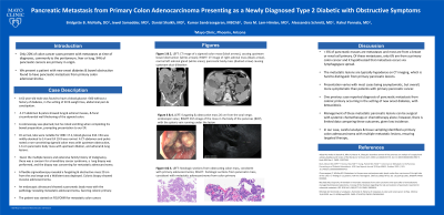
Figure: Figure 1: fungating & obstructive mass 20 cm from the anal verge, endoscopic view; Figure 2: EUS image of the mass in the body of the pancreas (BOP), with the splenic vein running under the lesion; Figure 3: CT image of Sigmoid colon mass (black arrows), causing upstream bowel obstruction (white arrows); Figure 4: CT image of right adrenal mass (black arrow), normal left adrenal gland (white areas), pancreatic body mass (dashed arrow) causing upstream duct dilatation; Figure 5: histologic sections from obstructing colon mass, consistent with primary adenocarcinoma; Figure 6: histologic sections from pancreatic mass, consistent with metastatic adenocarcinoma from colon primary
Disclosures:
Bridgette McNally indicated no relevant financial relationships.
Jewel Samadder: Caris Labs – Advisory Committee/Board Member. Recursion Pharma – Consultant.
Danial Shaikh indicated no relevant financial relationships.
Dora Lam-Himlin indicated no relevant financial relationships.
Alessandra Ceolin Schmitt indicated no relevant financial relationships.
Kumaresan Sandrasegaran indicated no relevant financial relationships.
Rahul Pannala: HCL Technologies – Consultant. Nestle Healthsciences – Advisor or Review Panel Member.
Bridgette B. McNally, DO1, Jewel Samadder, MD2, Danial Haris. Shaikh, MD3, Dora Lam-Himlin, MD4, Alessandra Ceolin Schmitt, MD4, Kumaresan Sandrasegaran, MBChB5, Rahul Pannala, MD4. P0322 - Pancreatic Metastasis from Primary Colon Adenocarcinoma Presenting as a Newly Diagnosed Type 2 Diabetic with Obstructive Symptoms, ACG 2024 Annual Scientific Meeting Abstracts. Philadelphia, PA: American College of Gastroenterology.
