Sunday Poster Session
Category: Colon
P0325 - Colonic Deception: Two Cases of Ischemic Colitis Posing as Malignant Masses
Sunday, October 27, 2024
3:30 PM - 7:00 PM ET
Location: Exhibit Hall E

Has Audio
- RS
Ruchi Sharma, MD
University of Iowa Hospitals & Clinics
Iowa City, IA
Presenting Author(s)
Vijayvardhan Kamalumpundi, BS1, Ruchi Sharma, MD2, Yiqin Xiong, MD, PhD2, Estefania M. Flores Cordova, MD3, Divya Ashat, MBBS2, Mohammad Ansari, MD2
1Roy J. and Lucille A. Carver College of Medicine, Iowa City, IA; 2University of Iowa Hospitals & Clinics, Iowa City, IA; 3University of Iowa Hospitals and Clinics, Coralville, IA
Introduction: Isolated right-sided colonic ischemia (IRCI) is a well-known entity, however, it occurs far less frequently than left-sided colonic ischemia. Scattered reports suggest that IRCI can mimic malignancy. We report two cases of ischemic colitis presenting as a cecal mass.
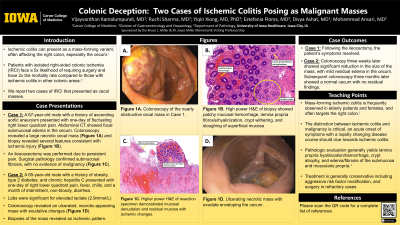
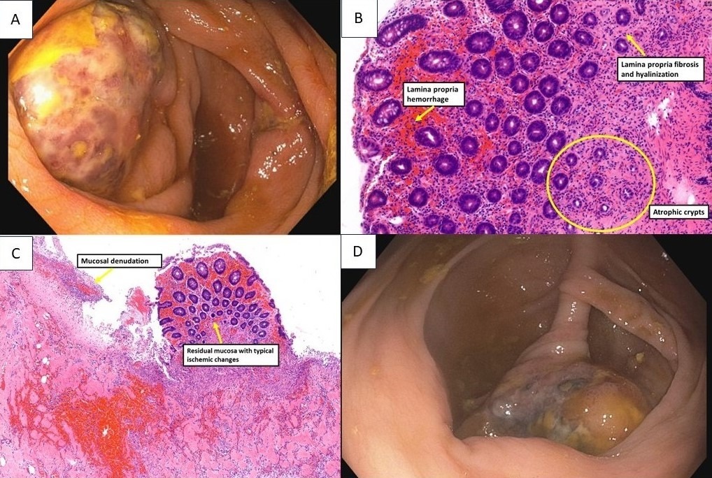
Case Description/Methods: A 67-year-old male with a history of ascending aortic aneurysm, known aortic clot, and diverticulosis presented to the Emergency Department (ED) with a 1-day history of fluctuating right lower quadrant pain. Labs were notable for mild leukocytosis. Abdominal CT revealed focal changes in the cecum including submucosal edema, decreased mucosal enhancement, and possible pneumatosis. Colonoscopy showed a large, necrotic, cecal mass (Figure 1A). Biopsies of the mass showed patchy mucosal hemorrhage, lamina propria fibrosis and hyalinization, crypt withering, and sloughing of the superficial mucosa, consistent with ischemic injury (Figure 1B). Due to ongoing abdominal pain, the patient underwent an ileocecectomy with resolution of abdominal pain. Surgical pathology showed marked submucosal fibrosis and edema, with no evidence of malignancy (Figure 1C).
A 65-year-old male with a history of obesity, type 2 diabetes, and chronic hepatitis C presented to the ED with a 1-day history of right lower quadrant pain, fever, chills, and a month of intermittent non-bloody diarrhea. Labs were significant for lactate 2.9 mmol/L. Enteric panel and C. difficile toxin were negative. CT abdomen showed focal inflammatory changes of the cecum. Colonoscopy revealed an ulcerated necrotic-appearing mass with exudate enveloping the cecum (Figure 1D). Final pathology revealed an acute ischemic pattern with no neoplastic cells. A colonoscopy three weeks later showed significant reduction in the size of the mass, with mild residual edema in the cecum. Subsequent colonoscopy three months later showed a normal cecum with no residual findings.
Discussion: IRCI can present as a mass due to submucosal hemorrhage and edema. Pathologic features include lamina propria hyalinization, hemorrhage, and loss of surface and upper crypt epithelium among others. Conservative management is generally first line treatment as patients with IRCI have higher surgical mortality. Our patients were successfully treated with surgical management and conservative treatment respectively. We report these cases to increase clinical awareness of IRCI presenting as a mass lesion to ensure accurate intervention for these patients.

Disclosures:
Vijayvardhan Kamalumpundi, BS1, Ruchi Sharma, MD2, Yiqin Xiong, MD, PhD2, Estefania M. Flores Cordova, MD3, Divya Ashat, MBBS2, Mohammad Ansari, MD2. P0325 - Colonic Deception: Two Cases of Ischemic Colitis Posing as Malignant Masses, ACG 2024 Annual Scientific Meeting Abstracts. Philadelphia, PA: American College of Gastroenterology.
1Roy J. and Lucille A. Carver College of Medicine, Iowa City, IA; 2University of Iowa Hospitals & Clinics, Iowa City, IA; 3University of Iowa Hospitals and Clinics, Coralville, IA
Introduction: Isolated right-sided colonic ischemia (IRCI) is a well-known entity, however, it occurs far less frequently than left-sided colonic ischemia. Scattered reports suggest that IRCI can mimic malignancy. We report two cases of ischemic colitis presenting as a cecal mass.
Case Description/Methods: A 67-year-old male with a history of ascending aortic aneurysm, known aortic clot, and diverticulosis presented to the Emergency Department (ED) with a 1-day history of fluctuating right lower quadrant pain. Labs were notable for mild leukocytosis. Abdominal CT revealed focal changes in the cecum including submucosal edema, decreased mucosal enhancement, and possible pneumatosis. Colonoscopy showed a large, necrotic, cecal mass (Figure 1A). Biopsies of the mass showed patchy mucosal hemorrhage, lamina propria fibrosis and hyalinization, crypt withering, and sloughing of the superficial mucosa, consistent with ischemic injury (Figure 1B). Due to ongoing abdominal pain, the patient underwent an ileocecectomy with resolution of abdominal pain. Surgical pathology showed marked submucosal fibrosis and edema, with no evidence of malignancy (Figure 1C).
A 65-year-old male with a history of obesity, type 2 diabetes, and chronic hepatitis C presented to the ED with a 1-day history of right lower quadrant pain, fever, chills, and a month of intermittent non-bloody diarrhea. Labs were significant for lactate 2.9 mmol/L. Enteric panel and C. difficile toxin were negative. CT abdomen showed focal inflammatory changes of the cecum. Colonoscopy revealed an ulcerated necrotic-appearing mass with exudate enveloping the cecum (Figure 1D). Final pathology revealed an acute ischemic pattern with no neoplastic cells. A colonoscopy three weeks later showed significant reduction in the size of the mass, with mild residual edema in the cecum. Subsequent colonoscopy three months later showed a normal cecum with no residual findings.
Discussion: IRCI can present as a mass due to submucosal hemorrhage and edema. Pathologic features include lamina propria hyalinization, hemorrhage, and loss of surface and upper crypt epithelium among others. Conservative management is generally first line treatment as patients with IRCI have higher surgical mortality. Our patients were successfully treated with surgical management and conservative treatment respectively. We report these cases to increase clinical awareness of IRCI presenting as a mass lesion to ensure accurate intervention for these patients.

Figure: Figure 1A: Colonoscopy of the nearly obstructive cecal mass in Case 1. Figure 1B: High power H&E stain of biopsy showed patchy mucosal hemorrhage, lamina propria fibrosis/hyalinization, crypt withering, and sloughing of superficial mucosa (yellow arrows). Figure 1C: Higher power H&E of resection specimen demonstrated mucosal denudation and residual mucosa with ischemic changes (yellow arrows). Figure 1D: Ulcerating necrotic mass with exudate enveloping the cecum.
Disclosures:
Vijayvardhan Kamalumpundi indicated no relevant financial relationships.
Ruchi Sharma indicated no relevant financial relationships.
Yiqin Xiong indicated no relevant financial relationships.
Estefania Flores Cordova indicated no relevant financial relationships.
Divya Ashat indicated no relevant financial relationships.
Mohammad Ansari indicated no relevant financial relationships.
Vijayvardhan Kamalumpundi, BS1, Ruchi Sharma, MD2, Yiqin Xiong, MD, PhD2, Estefania M. Flores Cordova, MD3, Divya Ashat, MBBS2, Mohammad Ansari, MD2. P0325 - Colonic Deception: Two Cases of Ischemic Colitis Posing as Malignant Masses, ACG 2024 Annual Scientific Meeting Abstracts. Philadelphia, PA: American College of Gastroenterology.
