Sunday Poster Session
Category: Colon
P0327 - Unmasking Epiploic Appendagitis: A Hidden Cause of Acute Abdominal Pain
Sunday, October 27, 2024
3:30 PM - 7:00 PM ET
Location: Exhibit Hall E

Has Audio
- AD
Aladdin Dahbour, MD
Indiana University
Evansville, IN
Presenting Author(s)
Aladdin Dahbour, MD1, Kyle Hagen, DO2, Oluwagbenga Serrano, MD, FACG3
1Indiana University, Evansville, IN; 2Indiana University School of Medicine, Evansville, IN; 3Good Samaritan Hospital, Vincennes, IN
Introduction: Epiploic appendagitis is characterized by inflammation of the epiploic appendages, which are small fat-filled pouches on the exterior of the colon. It often presents with localized abdominal pain and can be misdiagnosed due to its nonspecific presentation. This case highlights the importance of including epiploic appendagitis in the differential diagnosis of acute abdominal pain.
Case Description/Methods: A 48-year-old female presented to the emergency room with acute RUQ abdominal pain. Her medical history was unremarkable. On initial evaluation, her vitals were stable, and a CT abdomen without contrast showed no abnormalities. However, the following day, her pain significantly worsened, and she developed hypertension and a fever of 100.9°F.
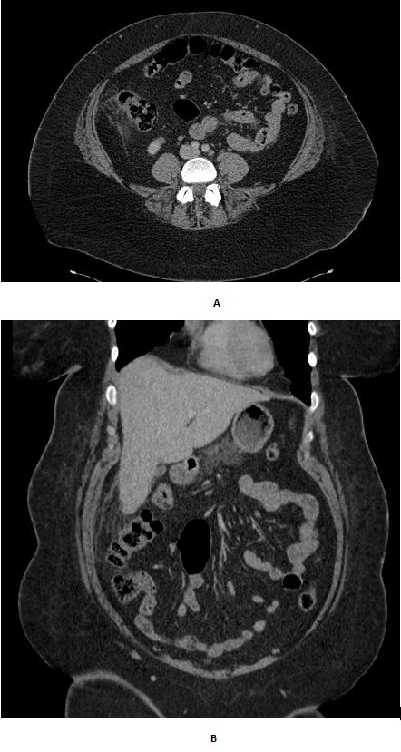
She returned to the ER, and a CT abdomen with contrast revealed nonspecific fat stranding along the epiploic fat of the ascending colon, consistent with epiploic appendagitis. Blood work and urinalysis were unremarkable.
The patient was treated with Naproxen and Metronidazole, showing significant improvement within five days.
Discussion: Epiploic appendagitis is a self-limiting condition that is often managed conservatively with anti-inflammatory medications and sometimes antibiotics. Accurate diagnosis primarily relies on imaging, where CT scans can reveal characteristic fat stranding. Misdiagnosis can lead to unnecessary surgical interventions due to its mimicry of other acute abdominal conditions such as appendicitis or diverticulitis.
The primary diagnostic tools for epiploic appendagitis are ultrasound (US) and computed tomography (CT), with CT being particularly valuable for its detailed imaging. CT typically shows a fat-density lesion, 1.5 to 3.5 cm in diameter, adjacent to the anterior colon wall, often the sigmoid colon or cecum. Magnetic resonance imaging (MRI) can also be useful, offering additional soft tissue contrast. Accurate imaging helps avoid unnecessary surgeries and ensures proper medical management.
Management of epiploic appendagitis is usually conservative, using pain control and anti-inflammatory medications. Surgery is rarely needed and reserved for diagnostic uncertainty or complications.
This case highlights the necessity of considering epiploic appendagitis in patients presenting with acute abdominal pain. Prompt imaging and conservative management can lead to rapid symptom resolution and prevent unnecessary surgical procedures.

Disclosures:
Aladdin Dahbour, MD1, Kyle Hagen, DO2, Oluwagbenga Serrano, MD, FACG3. P0327 - Unmasking Epiploic Appendagitis: A Hidden Cause of Acute Abdominal Pain, ACG 2024 Annual Scientific Meeting Abstracts. Philadelphia, PA: American College of Gastroenterology.
1Indiana University, Evansville, IN; 2Indiana University School of Medicine, Evansville, IN; 3Good Samaritan Hospital, Vincennes, IN
Introduction: Epiploic appendagitis is characterized by inflammation of the epiploic appendages, which are small fat-filled pouches on the exterior of the colon. It often presents with localized abdominal pain and can be misdiagnosed due to its nonspecific presentation. This case highlights the importance of including epiploic appendagitis in the differential diagnosis of acute abdominal pain.
Case Description/Methods: A 48-year-old female presented to the emergency room with acute RUQ abdominal pain. Her medical history was unremarkable. On initial evaluation, her vitals were stable, and a CT abdomen without contrast showed no abnormalities. However, the following day, her pain significantly worsened, and she developed hypertension and a fever of 100.9°F.
She returned to the ER, and a CT abdomen with contrast revealed nonspecific fat stranding along the epiploic fat of the ascending colon, consistent with epiploic appendagitis. Blood work and urinalysis were unremarkable.
The patient was treated with Naproxen and Metronidazole, showing significant improvement within five days.
Discussion: Epiploic appendagitis is a self-limiting condition that is often managed conservatively with anti-inflammatory medications and sometimes antibiotics. Accurate diagnosis primarily relies on imaging, where CT scans can reveal characteristic fat stranding. Misdiagnosis can lead to unnecessary surgical interventions due to its mimicry of other acute abdominal conditions such as appendicitis or diverticulitis.
The primary diagnostic tools for epiploic appendagitis are ultrasound (US) and computed tomography (CT), with CT being particularly valuable for its detailed imaging. CT typically shows a fat-density lesion, 1.5 to 3.5 cm in diameter, adjacent to the anterior colon wall, often the sigmoid colon or cecum. Magnetic resonance imaging (MRI) can also be useful, offering additional soft tissue contrast. Accurate imaging helps avoid unnecessary surgeries and ensures proper medical management.
Management of epiploic appendagitis is usually conservative, using pain control and anti-inflammatory medications. Surgery is rarely needed and reserved for diagnostic uncertainty or complications.
This case highlights the necessity of considering epiploic appendagitis in patients presenting with acute abdominal pain. Prompt imaging and conservative management can lead to rapid symptom resolution and prevent unnecessary surgical procedures.

Figure: A,B:Axial and Coronal section of contrasted Abdominal CT showing Fat stranding in the Ascending colon.
Disclosures:
Aladdin Dahbour indicated no relevant financial relationships.
Kyle Hagen indicated no relevant financial relationships.
Oluwagbenga Serrano: MERCK – Stock-publicly held company(excluding mutual/index funds).
Aladdin Dahbour, MD1, Kyle Hagen, DO2, Oluwagbenga Serrano, MD, FACG3. P0327 - Unmasking Epiploic Appendagitis: A Hidden Cause of Acute Abdominal Pain, ACG 2024 Annual Scientific Meeting Abstracts. Philadelphia, PA: American College of Gastroenterology.
