Sunday Poster Session
Category: Colon
P0330 - A Case of Mistaken Identity: Fibrillar Confused for Splenic Abscess
Sunday, October 27, 2024
3:30 PM - 7:00 PM ET
Location: Exhibit Hall E

Has Audio

Bobby Thomas, MD
Elmhurst Hospital Center / Icahn School of Medicine at Mount Sinai
New York, NY
Presenting Author(s)
Bobby Thomas, MD1, Nirali Sheth, MD2, Richard Mitchell, MD3, Maliyat Matin, MD3, Woo Suk Kim, DO3, Bibhuti Adhikari, MD3, Joshua Aron, MD2, Aaron Walfish, MD3
1Elmhurst Hospital Center / Icahn School of Medicine at Mount Sinai, New York, NY; 2Elmhurst Hospital Center / Icahn School of Medicine at Mount Sinai, Elmhurst, NY; 3Elmhurst Hospital Center / Icahn School of Medicine at Mount Sinai, Queens, NY
Introduction: Splenic abscesses are rare and are often associated with diseases such as infective endocarditis, malignancy, and gastrointestinal perforation. They present with abdominal pain, abdominal distention, and fever. Occasionally, patients may present as having the signs and symptoms of an abscess, when in reality other factors are mimicking an infection.
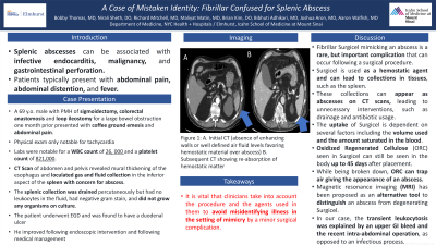
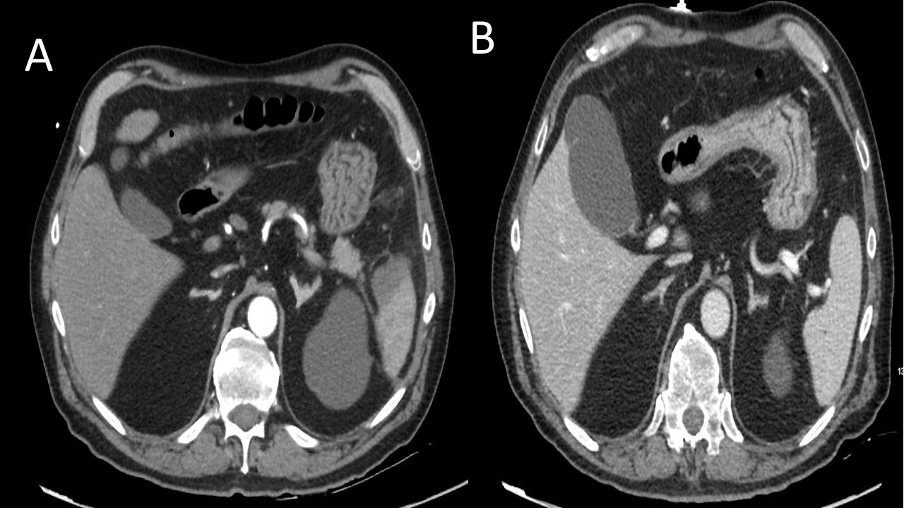
Case Description/Methods: A 69 year old male with a past medical history of sigmoidectomy, colorectal anastomosis and loop ileostomy from large bowel obstruction, and sigmoid stricture from diverticulum one month prior presented with coffee ground emesis and abdominal pain. His labs were notable for an elevated white blood cell count of 26, 000 and an elevated platelet count of 821,000. CT Angio Abdomen and Pelvis revealed mural thickening of the esophagus with concern for esophagitis and loculated gas and fluid collection in the inferior aspect of the spleen with concern for abscess. The splenic collection was drained by Interventional Radiology, but did not grow any organisms on culture, had negative gram stain, and had no leukocytes in the fluid. With leukocytosis secondary to hemoconcentration and negative cultures, the splenic collection was thought to be secondary to Fibrillar Surgicel rather than an abscess.
Discussion: Fibrillar Surgicel mimicking an abscess is a rare, but important complication that can occur following a surgical procedure. They are used as hemostatic agents and can lead to collections in tissues, such as the spleen. These collections can appear to be abscesses on CT scans, leading to unnecessary interventions, such as drainage and antibiotic usage. The uptake of Surgicel is dependent on several factors including the volume used and the amount saturated in the blood. Oxidized Regenerated Cellulose (ORC) seen in Surgicel can still be seen in the body up to 45 days after placement. While being broken down, ORC can trap air giving the appearance of an abscess. Magnetic resonance imaging (MRI) has been proposed as an alternative tool to distinguish an abscess from degenerating Surgicel. It is vital that clinicians and radiologists take into account the procedure and the agents used in them to avoid misidentifying illness in the setting of mimicry by a minor surgical complication. In our case, the transient leukocytosis was explained by an upper GI bleed and the recent intra-abdominal operation, as opposed to an infectious process.

Disclosures:
Bobby Thomas, MD1, Nirali Sheth, MD2, Richard Mitchell, MD3, Maliyat Matin, MD3, Woo Suk Kim, DO3, Bibhuti Adhikari, MD3, Joshua Aron, MD2, Aaron Walfish, MD3. P0330 - A Case of Mistaken Identity: Fibrillar Confused for Splenic Abscess, ACG 2024 Annual Scientific Meeting Abstracts. Philadelphia, PA: American College of Gastroenterology.
1Elmhurst Hospital Center / Icahn School of Medicine at Mount Sinai, New York, NY; 2Elmhurst Hospital Center / Icahn School of Medicine at Mount Sinai, Elmhurst, NY; 3Elmhurst Hospital Center / Icahn School of Medicine at Mount Sinai, Queens, NY
Introduction: Splenic abscesses are rare and are often associated with diseases such as infective endocarditis, malignancy, and gastrointestinal perforation. They present with abdominal pain, abdominal distention, and fever. Occasionally, patients may present as having the signs and symptoms of an abscess, when in reality other factors are mimicking an infection.
Case Description/Methods: A 69 year old male with a past medical history of sigmoidectomy, colorectal anastomosis and loop ileostomy from large bowel obstruction, and sigmoid stricture from diverticulum one month prior presented with coffee ground emesis and abdominal pain. His labs were notable for an elevated white blood cell count of 26, 000 and an elevated platelet count of 821,000. CT Angio Abdomen and Pelvis revealed mural thickening of the esophagus with concern for esophagitis and loculated gas and fluid collection in the inferior aspect of the spleen with concern for abscess. The splenic collection was drained by Interventional Radiology, but did not grow any organisms on culture, had negative gram stain, and had no leukocytes in the fluid. With leukocytosis secondary to hemoconcentration and negative cultures, the splenic collection was thought to be secondary to Fibrillar Surgicel rather than an abscess.
Discussion: Fibrillar Surgicel mimicking an abscess is a rare, but important complication that can occur following a surgical procedure. They are used as hemostatic agents and can lead to collections in tissues, such as the spleen. These collections can appear to be abscesses on CT scans, leading to unnecessary interventions, such as drainage and antibiotic usage. The uptake of Surgicel is dependent on several factors including the volume used and the amount saturated in the blood. Oxidized Regenerated Cellulose (ORC) seen in Surgicel can still be seen in the body up to 45 days after placement. While being broken down, ORC can trap air giving the appearance of an abscess. Magnetic resonance imaging (MRI) has been proposed as an alternative tool to distinguish an abscess from degenerating Surgicel. It is vital that clinicians and radiologists take into account the procedure and the agents used in them to avoid misidentifying illness in the setting of mimicry by a minor surgical complication. In our case, the transient leukocytosis was explained by an upper GI bleed and the recent intra-abdominal operation, as opposed to an infectious process.

Figure: Figure 1: A. Initial CT (absence of enhancing walls or well defined air fluid levels favoring hemostatic material over abscess) B. Subsequent CT showing re-absorption of hemostatic matter
Disclosures:
Bobby Thomas indicated no relevant financial relationships.
Nirali Sheth indicated no relevant financial relationships.
Richard Mitchell indicated no relevant financial relationships.
Maliyat Matin indicated no relevant financial relationships.
Woo Suk Kim indicated no relevant financial relationships.
Bibhuti Adhikari indicated no relevant financial relationships.
Joshua Aron indicated no relevant financial relationships.
Aaron Walfish indicated no relevant financial relationships.
Bobby Thomas, MD1, Nirali Sheth, MD2, Richard Mitchell, MD3, Maliyat Matin, MD3, Woo Suk Kim, DO3, Bibhuti Adhikari, MD3, Joshua Aron, MD2, Aaron Walfish, MD3. P0330 - A Case of Mistaken Identity: Fibrillar Confused for Splenic Abscess, ACG 2024 Annual Scientific Meeting Abstracts. Philadelphia, PA: American College of Gastroenterology.
