Sunday Poster Session
Category: Colon
P0376 - Serum β-hCG as a Prognostic Marker for Colorectal Adenocarcinoma in Women of Reproductive Age: A Case Report and Literature Review
Sunday, October 27, 2024
3:30 PM - 7:00 PM ET
Location: Exhibit Hall E

- CG
Changlin Gong, MD
NYC Health + Hospitals/Jacobi
Bronx, NY
Presenting Author(s)
Award: Presidential Poster Award
Changlin Gong, MD1, Fnu Vikash, MD, M.Med2, Juliet Silberstein, MD, MPH1, Sherrie White, MD1, Donald P. Kotler, MD3
1NYC Health + Hospitals/Jacobi, Bronx, NY; 2Albert Einstein College of Medicine, New York, NY; 3Albert Einstein College of Medicine, Bronx, NY
Introduction: Elevated serum levels of free β subunit of human chorionic gonadotropin (β-hCG) have been reported in 0-20% of patients with colorectal carcinoma. Histological examinations have also unveiled the presence of β-hCG in some cases of colorectal adenocarcinoma. Previously reported β-hCG-positive colon cancer were mostly identified in elderly patients. In females of reproductive age, positive serum β-hCG poses a unique challenge in differential diagnosis for clinicians but can serve as a tumor marker.
Case Description/Methods: A 30-year-old female was found to have adenocarcinoma of the sigmoid colon with liver metastasis, diagnosed by liver biopsy and mucosal biopsy under colonoscopy, which progressed very rapidly on imaging. On follow-up visits, β-hCG was tested due to complaints of abdominal pain, which turned out positive, raising concerns for pregnancy. Despite the absence of evidence for intrauterine or ectopic pregnancy on magnetic resonance imaging, transabdominal and transvaginal ultrasound, and curettage, β-hCG level continued to increase, reaching a peak of 3,556 U/L. Alternative etiologies of elevated β-hCG were explored, including tumor-related β-hCG production. Staining of biopsy samples came back positive for β-hCG on sigmoid colon mucosa but not on liver metastases. After administration of oxaliplatin, 5-fluorouracil, and leucovorin (modified FOLFOX-6) regimen, along with methotrexate, β-hCG level started to decrease.
Discussion: It was reported that β-hCG-positive colorectal adenocarcinoma is associated with worse survival, tissue invasion, and metastases, as well as good response to chemotherapy, compared to β-hCG-negative counterparts, which is similar to the behavior of trophoblastic tumors. Morphology of β-hCG-positive cells in colorectal adenocarcinoma on previous reports also hinted at the possibility of highly invasive, syncytiotrophoblast-like appearance and behavior of this specific subgroup of tumors. It was found that β-hCG-positive cells were more likely to be distributed at the periphery of the tumor or arranged in clumps resembling syncytiotrophoblasts, which could be a manifestation of de-differentiation of malignant tissue and could facilitate progression and metastasis. Moreover, serum β-hCG level decreases with antineoplastic treatment. Thus, β-hCG can serve as a tumor marker for response monitoring. However, more studies are needed to investigate the potential benefit of routine serological and histological β-hCG studies in patients with colorectal adenocarcinoma.

Disclosures:
Changlin Gong, MD1, Fnu Vikash, MD, M.Med2, Juliet Silberstein, MD, MPH1, Sherrie White, MD1, Donald P. Kotler, MD3. P0376 - Serum β-hCG as a Prognostic Marker for Colorectal Adenocarcinoma in Women of Reproductive Age: A Case Report and Literature Review, ACG 2024 Annual Scientific Meeting Abstracts. Philadelphia, PA: American College of Gastroenterology.
Changlin Gong, MD1, Fnu Vikash, MD, M.Med2, Juliet Silberstein, MD, MPH1, Sherrie White, MD1, Donald P. Kotler, MD3
1NYC Health + Hospitals/Jacobi, Bronx, NY; 2Albert Einstein College of Medicine, New York, NY; 3Albert Einstein College of Medicine, Bronx, NY
Introduction: Elevated serum levels of free β subunit of human chorionic gonadotropin (β-hCG) have been reported in 0-20% of patients with colorectal carcinoma. Histological examinations have also unveiled the presence of β-hCG in some cases of colorectal adenocarcinoma. Previously reported β-hCG-positive colon cancer were mostly identified in elderly patients. In females of reproductive age, positive serum β-hCG poses a unique challenge in differential diagnosis for clinicians but can serve as a tumor marker.
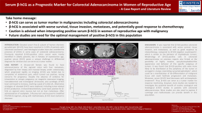
Case Description/Methods: A 30-year-old female was found to have adenocarcinoma of the sigmoid colon with liver metastasis, diagnosed by liver biopsy and mucosal biopsy under colonoscopy, which progressed very rapidly on imaging. On follow-up visits, β-hCG was tested due to complaints of abdominal pain, which turned out positive, raising concerns for pregnancy. Despite the absence of evidence for intrauterine or ectopic pregnancy on magnetic resonance imaging, transabdominal and transvaginal ultrasound, and curettage, β-hCG level continued to increase, reaching a peak of 3,556 U/L. Alternative etiologies of elevated β-hCG were explored, including tumor-related β-hCG production. Staining of biopsy samples came back positive for β-hCG on sigmoid colon mucosa but not on liver metastases. After administration of oxaliplatin, 5-fluorouracil, and leucovorin (modified FOLFOX-6) regimen, along with methotrexate, β-hCG level started to decrease.
Discussion: It was reported that β-hCG-positive colorectal adenocarcinoma is associated with worse survival, tissue invasion, and metastases, as well as good response to chemotherapy, compared to β-hCG-negative counterparts, which is similar to the behavior of trophoblastic tumors. Morphology of β-hCG-positive cells in colorectal adenocarcinoma on previous reports also hinted at the possibility of highly invasive, syncytiotrophoblast-like appearance and behavior of this specific subgroup of tumors. It was found that β-hCG-positive cells were more likely to be distributed at the periphery of the tumor or arranged in clumps resembling syncytiotrophoblasts, which could be a manifestation of de-differentiation of malignant tissue and could facilitate progression and metastasis. Moreover, serum β-hCG level decreases with antineoplastic treatment. Thus, β-hCG can serve as a tumor marker for response monitoring. However, more studies are needed to investigate the potential benefit of routine serological and histological β-hCG studies in patients with colorectal adenocarcinoma.

Figure: (A) Abdominal CT on first presentation showing mid-abdomen collection. (B) Abdominal CT on first presentation showing liver lesions. (C) Abdominal MRI one month later demonstrating worsened liver lesions. (D) H&E stain of biopsy sample 10×. (E) H&E stain of biopsy sample 40×. (F) β-hCG stain of biopsy sample. (G) Key events and changes in β-hCG level over time. CT, computed tomography; MRI, magnetic resonance imaging; H&E, hematoxylin and eosin; β-hCG, free β subunit of human chorionic gonadotropin; TAUS, transabdominal ultrasound; TVUS, transvaginal ultrasound; MTX, methotrexate.
Disclosures:
Changlin Gong indicated no relevant financial relationships.
Fnu Vikash indicated no relevant financial relationships.
Juliet Silberstein indicated no relevant financial relationships.
Sherrie White indicated no relevant financial relationships.
Donald Kotler: EMD Serono – Advisor or Review Panel Member.
Changlin Gong, MD1, Fnu Vikash, MD, M.Med2, Juliet Silberstein, MD, MPH1, Sherrie White, MD1, Donald P. Kotler, MD3. P0376 - Serum β-hCG as a Prognostic Marker for Colorectal Adenocarcinoma in Women of Reproductive Age: A Case Report and Literature Review, ACG 2024 Annual Scientific Meeting Abstracts. Philadelphia, PA: American College of Gastroenterology.


