Sunday Poster Session
Category: Colon
P0378 - Colonic Mantle Cell Lymphoma
Sunday, October 27, 2024
3:30 PM - 7:00 PM ET
Location: Exhibit Hall E

Has Audio
- SB
Sonya Bhaskar, MD
University of South Florida
Tampa, FL
Presenting Author(s)
Sonya Bhaskar, MD1, Jason Colizzo, MD2
1University of South Florida, Tampa, FL; 2James A. Haley Veterans' Hospital, Tampa, FL
Introduction: Mantle Cell Lymphoma (MCL) is a B-cell non-Hodgkin’s Lymphoma with a variable presentation and prognosis. In nodal MCL, gastrointestinal involvement is common and can involve any region. While common, endoscopic presentations can be variable, and here we present a case of incidental endoscopy findings in an asymptomatic patient.
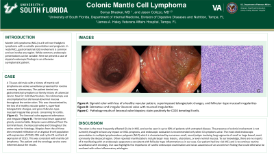
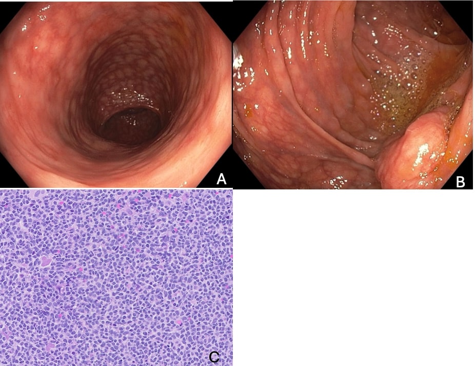
Case Description/Methods: A 73-year-old male with a history of mantle cell lymphoma on active surveillance presented for a screening colonoscopy. The patient denied any gastrointestinal symptoms or family history of colon cancer. His colonoscopy was uncomplicated and revealed mild diverticulosis and abnormal mucosa throughout the entire colon. The mucosa was characterized by the loss of a healthy vascular pattern, which superimposed telangiectatic changes, and follicular–type mucosal irregularities grossly which was concerning for colitis. (Figure A). The ileocecal valve appeared edematous and irregular (Figure B). The terminal ileum appeared grossly unremarkable. Biopsies were obtained from the terminal ileum, the ileocecal valve and throughout the entire colon for histology. Biopsies from the all three sites revealed infiltration of an atypical B-cell population with expression of CD20, CD5 and cyclin-D1 and lack of expression of CD23. This was consistent with mantle cell lymphoma. The patient and the oncology service were informed about the results.
Discussion: The colon is the most involved gastro-intestinal site in MCL and can be seen in up to 90% of patients with untreated disease. While involvement is common, gastro-intestinal involvement does not have clinical significance in prognosis and endoscopies are only recommended if the patient has symptoms. The most cited endoscopic presentation is multiple lymphomatous polyposis which is characterized by numerous, small, round polyps involving long segments of small or large bowel, most commonly the ileocecal region. Other reported manifestations include polyps, masses, and normal mucosa. To our knowledge, there are no reports of it manifesting with an appearance consistent with colitis as in our case. Our patient had a low risk MCL and thus the plan is for continued surveillance. Our case demonstrates the importance of a careful exam and the necessity of biopsies of abnormal findings.

Disclosures:
Sonya Bhaskar, MD1, Jason Colizzo, MD2. P0378 - Colonic Mantle Cell Lymphoma, ACG 2024 Annual Scientific Meeting Abstracts. Philadelphia, PA: American College of Gastroenterology.
1University of South Florida, Tampa, FL; 2James A. Haley Veterans' Hospital, Tampa, FL
Introduction: Mantle Cell Lymphoma (MCL) is a B-cell non-Hodgkin’s Lymphoma with a variable presentation and prognosis. In nodal MCL, gastrointestinal involvement is common and can involve any region. While common, endoscopic presentations can be variable, and here we present a case of incidental endoscopy findings in an asymptomatic patient.
Case Description/Methods: A 73-year-old male with a history of mantle cell lymphoma on active surveillance presented for a screening colonoscopy. The patient denied any gastrointestinal symptoms or family history of colon cancer. His colonoscopy was uncomplicated and revealed mild diverticulosis and abnormal mucosa throughout the entire colon. The mucosa was characterized by the loss of a healthy vascular pattern, which superimposed telangiectatic changes, and follicular–type mucosal irregularities grossly which was concerning for colitis. (Figure A). The ileocecal valve appeared edematous and irregular (Figure B). The terminal ileum appeared grossly unremarkable. Biopsies were obtained from the terminal ileum, the ileocecal valve and throughout the entire colon for histology. Biopsies from the all three sites revealed infiltration of an atypical B-cell population with expression of CD20, CD5 and cyclin-D1 and lack of expression of CD23. This was consistent with mantle cell lymphoma. The patient and the oncology service were informed about the results.
Discussion: The colon is the most involved gastro-intestinal site in MCL and can be seen in up to 90% of patients with untreated disease. While involvement is common, gastro-intestinal involvement does not have clinical significance in prognosis and endoscopies are only recommended if the patient has symptoms. The most cited endoscopic presentation is multiple lymphomatous polyposis which is characterized by numerous, small, round polyps involving long segments of small or large bowel, most commonly the ileocecal region. Other reported manifestations include polyps, masses, and normal mucosa. To our knowledge, there are no reports of it manifesting with an appearance consistent with colitis as in our case. Our patient had a low risk MCL and thus the plan is for continued surveillance. Our case demonstrates the importance of a careful exam and the necessity of biopsies of abnormal findings.

Figure: Figure A: Sigmoid colon with loss of a healthy vascular pattern, which superimposed telangiectatic changes, and follicular–type mucosal irregularities
Figure B: Edematous and irregular ileocecal valve with mucosal irregularities
Figure C: Biopsy of ileocecal valve stains positively for CD20 denoting that the cells are B-cells.
Figure B: Edematous and irregular ileocecal valve with mucosal irregularities
Figure C: Biopsy of ileocecal valve stains positively for CD20 denoting that the cells are B-cells.
Disclosures:
Sonya Bhaskar indicated no relevant financial relationships.
Jason Colizzo indicated no relevant financial relationships.
Sonya Bhaskar, MD1, Jason Colizzo, MD2. P0378 - Colonic Mantle Cell Lymphoma, ACG 2024 Annual Scientific Meeting Abstracts. Philadelphia, PA: American College of Gastroenterology.
