Sunday Poster Session
Category: Esophagus
P0569 - Scoping the Uncommon: Esophageal Epidermoid Metaplasia as a Premalignant Phenomenon
Sunday, October 27, 2024
3:30 PM - 7:00 PM ET
Location: Exhibit Hall E

Has Audio

Furqan A. Bhullar, MD
St. Joseph's University Medical Center
Presenting Author(s)
Furqan A. Bhullar, MD, Gabriel Melki, MD, Momina Sharif Umair, MD, Essam Ahmed, MD, Yana Cavanagh, MD
St. Joseph's University Medical Center, Paterson, NJ
Introduction: Esophageal cancer ranks as the eighth most frequently diagnosed cancer globally and stands as the sixth leading cause of cancer-related deaths. Esophageal cancer is mainly divided into two subtypes: adenocarcinoma, dominant in developed nations, and squamous cell carcinoma (ESCC), more prevalent in developing nations, making up 80-90% of global cases. Esophageal epidermoid metaplasia (EEM) is a rare condition, potentially premalignant, associated with squamous cell carcinoma (ESCC). To the best of our knowledge, this entity was first described in 1997 by Nakanishi and colleagues where they describe white, well-demarcated areas within the distal esophagus regions exhibiting hyperorthokeratosis and hypergranulosis, a condition referred to as 'epidermization'. Investigative efforts to quantify the incidence of EEM are notably infrequent, leading to potentially skewed prevalence data, confounded by the absence of a universally accepted pathological definition. This case report addresses the incidental finding of EEM during endoscopy and its clinical and histopathological characteristics.
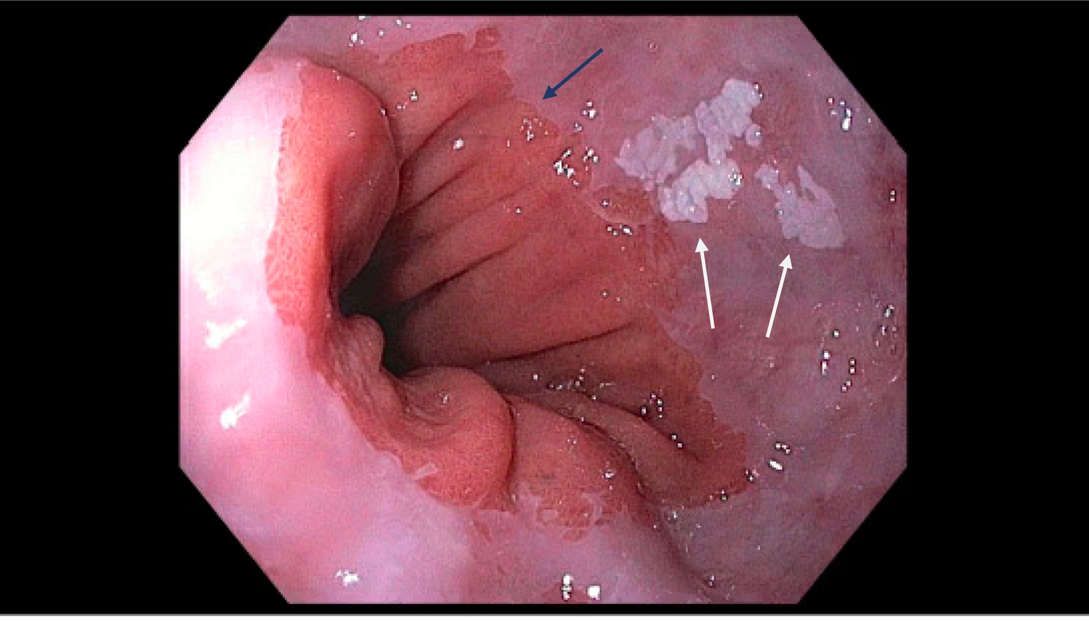
Case Description/Methods: We report a case of a 53-year-old male with significant medical history including chronic pancreatitis, presenting with epigastric pain. An incidental finding of a white esophageal plaque during an endoscopy conducted to evaluate a pancreatic mass was biopsied with cold forceps. Pathology of the distal esophageal plaque biopsy revealed focal hyperkeratosis, parakeratosis and hypergranulosis indicating benign esophageal mucosa with focal epidermoid metaplasia suggesting EEM, an observation that adds to the scant literature on the condition. There was no significant inflammation or fungal organisms identified.
Discussion: ESCC is a globally prevalent cancer with poor prognosis. EEM, while rare, has been implicated as a precursor to ESCC. Identified by distinctive endoscopic appearance and absence of Lugol's iodine staining, EEM's diagnosis underscores the need for meticulous endoscopic evaluation. Genetic studies highlight mutations common to both EEM and ESCC, suggesting a complex pathogenesis. The case emphasizes the importance of recognizing EEM's clinical presentation and the necessity for further research into its genetics and screening strategies, which could enhance early detection and intervention for ESCC. This report also suggests that the incidence of EEM may be underreported due to the absence of a universal pathological definition and standardized screening protocols.

Disclosures:
Furqan A. Bhullar, MD, Gabriel Melki, MD, Momina Sharif Umair, MD, Essam Ahmed, MD, Yana Cavanagh, MD. P0569 - Scoping the Uncommon: Esophageal Epidermoid Metaplasia as a Premalignant Phenomenon, ACG 2024 Annual Scientific Meeting Abstracts. Philadelphia, PA: American College of Gastroenterology.
St. Joseph's University Medical Center, Paterson, NJ
Introduction: Esophageal cancer ranks as the eighth most frequently diagnosed cancer globally and stands as the sixth leading cause of cancer-related deaths. Esophageal cancer is mainly divided into two subtypes: adenocarcinoma, dominant in developed nations, and squamous cell carcinoma (ESCC), more prevalent in developing nations, making up 80-90% of global cases. Esophageal epidermoid metaplasia (EEM) is a rare condition, potentially premalignant, associated with squamous cell carcinoma (ESCC). To the best of our knowledge, this entity was first described in 1997 by Nakanishi and colleagues where they describe white, well-demarcated areas within the distal esophagus regions exhibiting hyperorthokeratosis and hypergranulosis, a condition referred to as 'epidermization'. Investigative efforts to quantify the incidence of EEM are notably infrequent, leading to potentially skewed prevalence data, confounded by the absence of a universally accepted pathological definition. This case report addresses the incidental finding of EEM during endoscopy and its clinical and histopathological characteristics.
Case Description/Methods: We report a case of a 53-year-old male with significant medical history including chronic pancreatitis, presenting with epigastric pain. An incidental finding of a white esophageal plaque during an endoscopy conducted to evaluate a pancreatic mass was biopsied with cold forceps. Pathology of the distal esophageal plaque biopsy revealed focal hyperkeratosis, parakeratosis and hypergranulosis indicating benign esophageal mucosa with focal epidermoid metaplasia suggesting EEM, an observation that adds to the scant literature on the condition. There was no significant inflammation or fungal organisms identified.
Discussion: ESCC is a globally prevalent cancer with poor prognosis. EEM, while rare, has been implicated as a precursor to ESCC. Identified by distinctive endoscopic appearance and absence of Lugol's iodine staining, EEM's diagnosis underscores the need for meticulous endoscopic evaluation. Genetic studies highlight mutations common to both EEM and ESCC, suggesting a complex pathogenesis. The case emphasizes the importance of recognizing EEM's clinical presentation and the necessity for further research into its genetics and screening strategies, which could enhance early detection and intervention for ESCC. This report also suggests that the incidence of EEM may be underreported due to the absence of a universal pathological definition and standardized screening protocols.

Figure: Leukoplakia (white arrows) incidentally noted at the distal esophagus above the gastroesophageal junction (blue arrow) measuring 15mm in maximum diameter
Disclosures:
Furqan Bhullar indicated no relevant financial relationships.
Gabriel Melki indicated no relevant financial relationships.
Momina Sharif Umair indicated no relevant financial relationships.
Essam Ahmed indicated no relevant financial relationships.
Yana Cavanagh indicated no relevant financial relationships.
Furqan A. Bhullar, MD, Gabriel Melki, MD, Momina Sharif Umair, MD, Essam Ahmed, MD, Yana Cavanagh, MD. P0569 - Scoping the Uncommon: Esophageal Epidermoid Metaplasia as a Premalignant Phenomenon, ACG 2024 Annual Scientific Meeting Abstracts. Philadelphia, PA: American College of Gastroenterology.
