Sunday Poster Session
Category: Esophagus
P0574 - A Case of “Black Esophagus” Following Esophageal Candidiasis
Sunday, October 27, 2024
3:30 PM - 7:00 PM ET
Location: Exhibit Hall E

Has Audio

Kemmian D. Johnson, MD, MPH
University of Arkansas for Medical Sciences
Little Rock, AR
Presenting Author(s)
Kemmian D. Johnson, MD, MPH, Genesis Perez Del Nogal, MD, Meer Akbar. Ali, MD, Camila Simoes, MD
University of Arkansas for Medical Sciences, Little Rock, AR
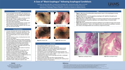
Introduction: Acute esophageal necrosis (AEN), or “black esophagus”, is a rare condition characterized by the endoscopic appearance of diffuse black coloration of the esophageal mucosa. The incidence is higher in older individuals, with a predilection for males. The commonest presenting symptoms include hematemesis and melena. The etiology of AEN remains unclear. However, several factors have been implicated in its development, including hypoperfusion, antibiotics, alcoholic hepatitis, diabetic ketoacidosis and underlying malignancy. Additionally, viral and fungal infections have been reported. Herein, we present a rare case of esophageal necrosis with concomitant candida esophagitis.
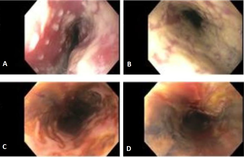
Case Description/Methods: A 43 year old woman with common variable immune deficiency, deficiency of adenosine deaminase 2, presented with fever, lymphadenopathy, and an absolute neutrophil count of 0. Her infectious workup, including blood and urine cultures, was negative. She was treated with Cefepime for febrile neutropenia. During her stay, the patient reported odynophagia following ingestion of a potassium pill. An Esophagogastroduodenoscopy (EGD) showed esophageal candidiasis, and confluent esophageal ulceration from the upper esophageal sphincter to the mid esophagus (Images A/B). Biopsies reported multiple large clusters of numerous fungal species. Subsequently, a repeat EGD with cultures was performed five days later, which revealed a necrotic-appearing esophagus with significant sloughing and ulceration, which raised concerns for liquefactive necrosis (Images C/D). Ultimately, repeat biopsies were not obtained due to the poor integrity of the esophageal mucosa. Thoracic surgery was consulted, but the patient ultimately declined surgery and was started on total parenteral nutrition (TPN).
Discussion: The prevalence of AEN is less than 0.3% based on documented reports and autopsies. While any portion of the esophagus can be affected, the distal esophagus is most commonly involved. Here we present a case of a young immunocompromised female patient with extensive AED from the upper to the distal esophagus who presented with rapid onset dysphagia and odynophagia. Supportive management, treatment of underlying causes, intravenous proton pump inhibitors, and TPN for malnourished patients can lead to endoscopic resolution in most cases. Unfortunately, our patient’s poor prognosis precluded further endoscopic procedures, which were deemed to have no significant impact on her outcome.

Disclosures:
Kemmian D. Johnson, MD, MPH, Genesis Perez Del Nogal, MD, Meer Akbar. Ali, MD, Camila Simoes, MD. P0574 - A Case of “Black Esophagus” Following Esophageal Candidiasis, ACG 2024 Annual Scientific Meeting Abstracts. Philadelphia, PA: American College of Gastroenterology.
University of Arkansas for Medical Sciences, Little Rock, AR
Introduction: Acute esophageal necrosis (AEN), or “black esophagus”, is a rare condition characterized by the endoscopic appearance of diffuse black coloration of the esophageal mucosa. The incidence is higher in older individuals, with a predilection for males. The commonest presenting symptoms include hematemesis and melena. The etiology of AEN remains unclear. However, several factors have been implicated in its development, including hypoperfusion, antibiotics, alcoholic hepatitis, diabetic ketoacidosis and underlying malignancy. Additionally, viral and fungal infections have been reported. Herein, we present a rare case of esophageal necrosis with concomitant candida esophagitis.
Case Description/Methods: A 43 year old woman with common variable immune deficiency, deficiency of adenosine deaminase 2, presented with fever, lymphadenopathy, and an absolute neutrophil count of 0. Her infectious workup, including blood and urine cultures, was negative. She was treated with Cefepime for febrile neutropenia. During her stay, the patient reported odynophagia following ingestion of a potassium pill. An Esophagogastroduodenoscopy (EGD) showed esophageal candidiasis, and confluent esophageal ulceration from the upper esophageal sphincter to the mid esophagus (Images A/B). Biopsies reported multiple large clusters of numerous fungal species. Subsequently, a repeat EGD with cultures was performed five days later, which revealed a necrotic-appearing esophagus with significant sloughing and ulceration, which raised concerns for liquefactive necrosis (Images C/D). Ultimately, repeat biopsies were not obtained due to the poor integrity of the esophageal mucosa. Thoracic surgery was consulted, but the patient ultimately declined surgery and was started on total parenteral nutrition (TPN).
Discussion: The prevalence of AEN is less than 0.3% based on documented reports and autopsies. While any portion of the esophagus can be affected, the distal esophagus is most commonly involved. Here we present a case of a young immunocompromised female patient with extensive AED from the upper to the distal esophagus who presented with rapid onset dysphagia and odynophagia. Supportive management, treatment of underlying causes, intravenous proton pump inhibitors, and TPN for malnourished patients can lead to endoscopic resolution in most cases. Unfortunately, our patient’s poor prognosis precluded further endoscopic procedures, which were deemed to have no significant impact on her outcome.

Figure: Figures A/B. Esophageal candidiasis. Confluent esophageal ulceration from the upper esophageal sphincter to the mid esophagus.
Figures C/D. Necrotic appearing esophagus with significant sloughing and ulceration.
Figures C/D. Necrotic appearing esophagus with significant sloughing and ulceration.
Disclosures:
Kemmian Johnson indicated no relevant financial relationships.
Genesis Perez Del Nogal indicated no relevant financial relationships.
Meer Ali indicated no relevant financial relationships.
Camila Simoes indicated no relevant financial relationships.
Kemmian D. Johnson, MD, MPH, Genesis Perez Del Nogal, MD, Meer Akbar. Ali, MD, Camila Simoes, MD. P0574 - A Case of “Black Esophagus” Following Esophageal Candidiasis, ACG 2024 Annual Scientific Meeting Abstracts. Philadelphia, PA: American College of Gastroenterology.
