Sunday Poster Session
Category: Esophagus
P0604 - Esophageal Perforation Resulting From Nonaccidental Trauma in a Neonate
Sunday, October 27, 2024
3:30 PM - 7:00 PM ET
Location: Exhibit Hall E

Has Audio

Lanah Almatroud, BA
Michigan State University College of Human Medicine
Ann Arbor, MI
Presenting Author(s)
Lanah Almatroud, BA1, Jacob Hesen, 2, Judy Teran, BS3, Rashed Hasan, MD4
1Michigan State University College of Human Medicine, Ann Arbor, MI; 2University of Michigan, Ann Arbor, MI; 3Michigan State University College of Human Medicine, Novi, MI; 4Hurley Medical Center, Flint, MI
Introduction: Esophageal perforation (EP) due to nonaccidental trauma in neonates is extremely rare. We report a neonate with stridor, respiratory distress, and bloody vomitus. Radiographic and endoscopic evaluations were consistent with EP. To our knowledge, this is the youngest reported case of EP due to nonaccidental trauma.
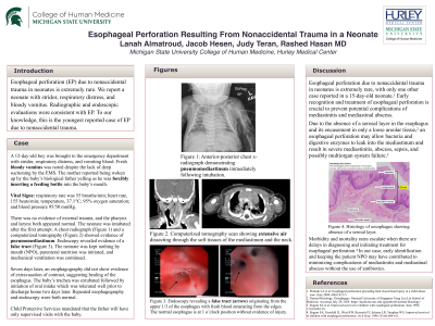
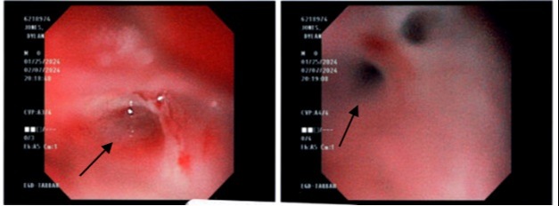
Case Description/Methods: A 12-day-old boy was brought to the emergency department with stridor, respiratory distress, and vomiting blood; respiratory rate was 55 breaths/min; heart rate, 155 beats/min; temperature, 37.1°C; and 95% oxygen saturation. Fresh bloody vomitus was noted despite the lack of deep suctioning or instrumentation by the EMS. The mother reported being woken up by the baby’s father yelling as he was forcibly inserting a feeding bottle into the baby’s mouth. There was no evidence of external trauma. The neonate was intubated after the first attempt, and the pharynx and larynx appeared normal. The chest radiograph following intubation showed evidence of pneumomediastinum. Computerized Tomography showed extensive air dissecting through the soft tissues of the mediastinum and the neck. Endoscopy revealed a false tract originating from the upper ⅓ of the esophagus in addition to the normal esophageal opening (Figure 1). The neonate was kept nothing by mouth (NPO), on mechanical ventilation, and received parenteral nutrition. CPS recommended that the father will have only supervised visits with the baby. Seven days later, an esophagography did not show evidence of extravasation of contrast. The baby’s trachea was extubated, followed by initiation of oral intake, which was tolerated well before discharge with the mother. Repeated esophagography and endoscopy were both normal.
Discussion: Only one other case of EP due to nonaccidental trauma in a 15-day-old neonate has been reported.1 Early recognition and treatment of EP is crucial to prevent mediastinitis and mediastinal abscess. Due to the absence of a serosal layer in the esophagus, an EP allows bacteria and digestive enzymes to leak into the mediastinum and may result in mediastinitis, abscess, sepsis, and possibly multiorgan system failure.2 In our case, early identification and keeping the patient NPO may have contributed to minimizing complications of mediastinitis and mediastinal abscess.
1. Pramuk LA et al. Esophageal perforation preceding fatal closed head injury in a child abuse case. 2004 Jun; 68(6):831-5.
2. Engum SA et al. Improved survival in children with esophageal perforation.1996 Jun; 131(6):604-10.

Disclosures:
Lanah Almatroud, BA1, Jacob Hesen, 2, Judy Teran, BS3, Rashed Hasan, MD4. P0604 - Esophageal Perforation Resulting From Nonaccidental Trauma in a Neonate, ACG 2024 Annual Scientific Meeting Abstracts. Philadelphia, PA: American College of Gastroenterology.
1Michigan State University College of Human Medicine, Ann Arbor, MI; 2University of Michigan, Ann Arbor, MI; 3Michigan State University College of Human Medicine, Novi, MI; 4Hurley Medical Center, Flint, MI
Introduction: Esophageal perforation (EP) due to nonaccidental trauma in neonates is extremely rare. We report a neonate with stridor, respiratory distress, and bloody vomitus. Radiographic and endoscopic evaluations were consistent with EP. To our knowledge, this is the youngest reported case of EP due to nonaccidental trauma.
Case Description/Methods: A 12-day-old boy was brought to the emergency department with stridor, respiratory distress, and vomiting blood; respiratory rate was 55 breaths/min; heart rate, 155 beats/min; temperature, 37.1°C; and 95% oxygen saturation. Fresh bloody vomitus was noted despite the lack of deep suctioning or instrumentation by the EMS. The mother reported being woken up by the baby’s father yelling as he was forcibly inserting a feeding bottle into the baby’s mouth. There was no evidence of external trauma. The neonate was intubated after the first attempt, and the pharynx and larynx appeared normal. The chest radiograph following intubation showed evidence of pneumomediastinum. Computerized Tomography showed extensive air dissecting through the soft tissues of the mediastinum and the neck. Endoscopy revealed a false tract originating from the upper ⅓ of the esophagus in addition to the normal esophageal opening (Figure 1). The neonate was kept nothing by mouth (NPO), on mechanical ventilation, and received parenteral nutrition. CPS recommended that the father will have only supervised visits with the baby. Seven days later, an esophagography did not show evidence of extravasation of contrast. The baby’s trachea was extubated, followed by initiation of oral intake, which was tolerated well before discharge with the mother. Repeated esophagography and endoscopy were both normal.
Discussion: Only one other case of EP due to nonaccidental trauma in a 15-day-old neonate has been reported.1 Early recognition and treatment of EP is crucial to prevent mediastinitis and mediastinal abscess. Due to the absence of a serosal layer in the esophagus, an EP allows bacteria and digestive enzymes to leak into the mediastinum and may result in mediastinitis, abscess, sepsis, and possibly multiorgan system failure.2 In our case, early identification and keeping the patient NPO may have contributed to minimizing complications of mediastinitis and mediastinal abscess.
1. Pramuk LA et al. Esophageal perforation preceding fatal closed head injury in a child abuse case. 2004 Jun; 68(6):831-5.
2. Engum SA et al. Improved survival in children with esophageal perforation.1996 Jun; 131(6):604-10.

Figure: Figure 1. Endoscopy revealing a false tract (arrow) with fresh blood emanating from the edges. The normal esophagus at the 1 o’clock position without evidence of injury at the edges.
Disclosures:
Lanah Almatroud indicated no relevant financial relationships.
Jacob Hesen indicated no relevant financial relationships.
Judy Teran indicated no relevant financial relationships.
Rashed Hasan indicated no relevant financial relationships.
Lanah Almatroud, BA1, Jacob Hesen, 2, Judy Teran, BS3, Rashed Hasan, MD4. P0604 - Esophageal Perforation Resulting From Nonaccidental Trauma in a Neonate, ACG 2024 Annual Scientific Meeting Abstracts. Philadelphia, PA: American College of Gastroenterology.
