Sunday Poster Session
Category: General Endoscopy
P0693 - An UncomMEN Cause of Jaundice
Sunday, October 27, 2024
3:30 PM - 7:00 PM ET
Location: Exhibit Hall E

Has Audio

Karthikeyan Baskaran, DO
Memorial Health University Medical Center
Savannah, GA
Presenting Author(s)
Karthikeyan Baskaran, DO, Victoria Grumbles, MD, Jonathan Kandiah, MD
Memorial Health University Medical Center, Savannah, GA
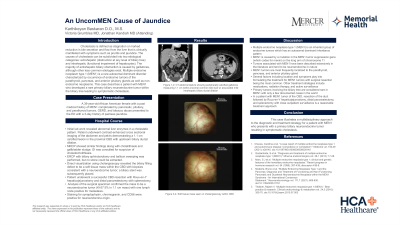
Introduction: Cholestasis is defined as reduction in bile flow from the liver which leads to symptoms such as pruritus and jaundice. The causes of cholestasis can be subdivided into two etiological categories: extrahepatic and intrahepatic. The majority of extrahepatic biliary obstruction is caused by gallstones, although other less common etiologies exist. Multiple endocrine neoplasm type 1 (MEN1) is a rare inherited autosomal dominant disorder characterized by the occurrence of endocrine tumors of the parathyroid, pancreas and anterior pituitary glands as well as non-endocrine neoplasms. This case describes a patient with MEN1 who developed a rare primary biliary neuroendocrine tumor within the biliary tree leading to symptomatic cholestasis.
Case Description/Methods: A 39-year old female with a past medical history of MEN1 complicated by pancreatic, pituitary and parathyroid tumors presented to the ED with a three-day history of painless jaundice. Her workup revealed abnormal liver enzymes in a cholestatic pattern. She underwent CT abdomen/pelvis that demonstrated a calcified lesion measuring 1.1 cm in the proximal common bile duct with upstream biliary ductal dilatation. MRCP showed similar findings along with cholelithiasis and gallbladder sludge. Gastroenterology was consulted for suspicion of choledocholithiasis. ERCP with biliary sphincterotomy and balloon sweeping was performed but no stone could be extracted. Further evaluation using cholangioscopy revealed the biliary filling defect to be a soft tissue mass within the common bile duct. Biopsies of the mass were consistent with a neuroendocrine tumor (NET). A biliary stent was placed and was referred for surgical excision.
She underwent common bile duct resection with Roux-en-Y hepaticojejunostomy and distal pancreatectomy with splenectomy. Analysis of the surgical specimens confirmed the bile duct mass to be a neuroendocrine tumor (Ki-57 5% in 1.1 cm mass) with one lymph node positive for metastatic tumor. Staining for synaptophysin, chromogranin and CD56 were positive, consistent with a neuroendocrine origin. The patient was discharged with plans for surveillance imaging.
Discussion: Tumors associated with MEN1 have been described extensively in the literature. Primary tumors involving the biliary tree are rare in MEN1 with few documented cases. This case illustrates a multidisciplinary approach to the diagnostic and treatment strategy for a patient with MEN1 who presents with a primary biliary NET resulting in symptomatic cholestasis.

Disclosures:
Karthikeyan Baskaran, DO, Victoria Grumbles, MD, Jonathan Kandiah, MD. P0693 - An UncomMEN Cause of Jaundice, ACG 2024 Annual Scientific Meeting Abstracts. Philadelphia, PA: American College of Gastroenterology.
Memorial Health University Medical Center, Savannah, GA
Introduction: Cholestasis is defined as reduction in bile flow from the liver which leads to symptoms such as pruritus and jaundice. The causes of cholestasis can be subdivided into two etiological categories: extrahepatic and intrahepatic. The majority of extrahepatic biliary obstruction is caused by gallstones, although other less common etiologies exist. Multiple endocrine neoplasm type 1 (MEN1) is a rare inherited autosomal dominant disorder characterized by the occurrence of endocrine tumors of the parathyroid, pancreas and anterior pituitary glands as well as non-endocrine neoplasms. This case describes a patient with MEN1 who developed a rare primary biliary neuroendocrine tumor within the biliary tree leading to symptomatic cholestasis.
Case Description/Methods: A 39-year old female with a past medical history of MEN1 complicated by pancreatic, pituitary and parathyroid tumors presented to the ED with a three-day history of painless jaundice. Her workup revealed abnormal liver enzymes in a cholestatic pattern. She underwent CT abdomen/pelvis that demonstrated a calcified lesion measuring 1.1 cm in the proximal common bile duct with upstream biliary ductal dilatation. MRCP showed similar findings along with cholelithiasis and gallbladder sludge. Gastroenterology was consulted for suspicion of choledocholithiasis. ERCP with biliary sphincterotomy and balloon sweeping was performed but no stone could be extracted. Further evaluation using cholangioscopy revealed the biliary filling defect to be a soft tissue mass within the common bile duct. Biopsies of the mass were consistent with a neuroendocrine tumor (NET). A biliary stent was placed and was referred for surgical excision.
She underwent common bile duct resection with Roux-en-Y hepaticojejunostomy and distal pancreatectomy with splenectomy. Analysis of the surgical specimens confirmed the bile duct mass to be a neuroendocrine tumor (Ki-57 5% in 1.1 cm mass) with one lymph node positive for metastatic tumor. Staining for synaptophysin, chromogranin and CD56 were positive, consistent with a neuroendocrine origin. The patient was discharged with plans for surveillance imaging.
Discussion: Tumors associated with MEN1 have been described extensively in the literature. Primary tumors involving the biliary tree are rare in MEN1 with few documented cases. This case illustrates a multidisciplinary approach to the diagnostic and treatment strategy for a patient with MEN1 who presents with a primary biliary NET resulting in symptomatic cholestasis.

Figure: Figure 1-2: CT abdomen/pelvis showing peripherally calcified gallstone measuring 1.1 cm within proximal common bile duct w/ associated mild intrahepatic biliary ductal dilation.
Figure 3-4: Soft tissue mass seen on cholangioscopy within CBD
Figure 3-4: Soft tissue mass seen on cholangioscopy within CBD
Disclosures:
Karthikeyan Baskaran indicated no relevant financial relationships.
Victoria Grumbles indicated no relevant financial relationships.
Jonathan Kandiah indicated no relevant financial relationships.
Karthikeyan Baskaran, DO, Victoria Grumbles, MD, Jonathan Kandiah, MD. P0693 - An UncomMEN Cause of Jaundice, ACG 2024 Annual Scientific Meeting Abstracts. Philadelphia, PA: American College of Gastroenterology.
