Sunday Poster Session
Category: GI Bleeding
P0769 - A Rare Case of Gastric Submucosal Lipoma Presenting With Upper GI bleed
Sunday, October 27, 2024
3:30 PM - 7:00 PM ET
Location: Exhibit Hall E

Has Audio
- SN
Sharanya Reddy Nemakallu, MBBS
Brookdale University Hospital Medical Center
Brooklyn, NY
Presenting Author(s)
Sharanya Reddy Nemakallu, MBBS1, Nader Daoud, MD2, Yasir Ali, MD2, Nicholas Paul Ridout, MD3, Derrick Cheung, MD2
1Brookdale University Hospital Medical Center, Brooklyn, NY; 2One Brooklyn Health-Brookdale University Hospital Medical Center, Brooklyn, NY; 3SUNY Downstate Medical Center, Brooklyn, NY
Introduction: Gastric lipomas are uncommon benign tumors typically discovered incidentally during upper endoscopic procedures or CT imaging. They comprise only 2-3% of all benign gastric tumors and about 5% of GI tract lipomas . They are predominantly located in the submucosa, often found in the pyloric antrum. While most gastric lipomas are asymptomatic, larger lesions can become symptomatic and present with obstruction or upper GI hemorrhage from ulceration.
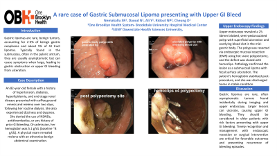
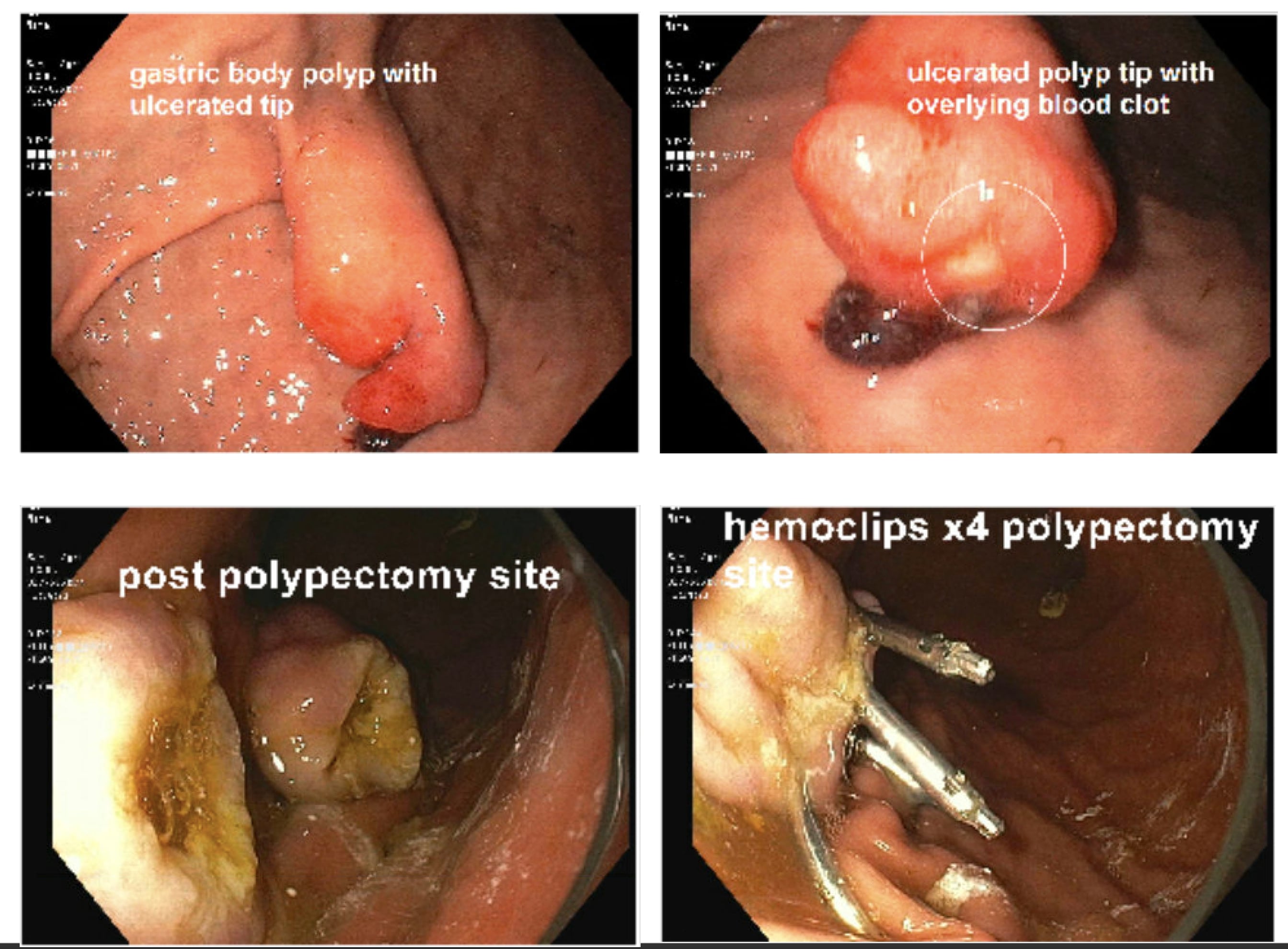
Case Description/Methods: An 82-year-old female with a medical history significant for hypertension, diabetes mellitus, hyperlipidemia, and end-stage renal disease presented to the hospital with multiple episodes of coffee ground emesis and black tarry stools over the past two days. Symptoms started after her routine dialysis session. She also reported dizziness and dyspnea on exertion since the morning of presentation. She denied the use of over-the-counter pain medications, NSAIDs, or any other antithrombotic agents. There was no history of similar episodes in the past. On admission, she was hemodynamically stable with a hemoglobin level of 5.1 g/dL (baseline ~8 g/dL). Physical examination revealed a soft, non-distended abdomen, non-tender on palpation, and rectal examination was positive for melena. Upper endoscopy performed during admission identified an ulcerated 25 mm semi-pedunculated polyp along the anterior wall of the mid gastric body, likely responsible for the patient's bleeding. Endoscopic mucosal resection (EMR) of the polyp was carried out using a hot snare polypectomy, with closure of the mucosal defect using 4 hemoclips. Pathological examination of the resected lesion confirmed it as a submucosal lipoma with focal surface ulceration. The patient's hemoglobin stabilized post-procedure, and she was discharged home.
Discussion: Gastric lipomas are rare, often asymptomatic tumors found incidentally during imaging and upper endoscopy. Larger lesions can ulcerate, causing upper GI bleeding. They should be considered in older patients with GI risk factors presenting with upper GI bleeding. Timely recognition and management with endoscopic resection or surgical intervention are critical for favorable outcomes and preventing recurrence of bleeding episodes.

Disclosures:
Sharanya Reddy Nemakallu, MBBS1, Nader Daoud, MD2, Yasir Ali, MD2, Nicholas Paul Ridout, MD3, Derrick Cheung, MD2. P0769 - A Rare Case of Gastric Submucosal Lipoma Presenting With Upper GI bleed, ACG 2024 Annual Scientific Meeting Abstracts. Philadelphia, PA: American College of Gastroenterology.
1Brookdale University Hospital Medical Center, Brooklyn, NY; 2One Brooklyn Health-Brookdale University Hospital Medical Center, Brooklyn, NY; 3SUNY Downstate Medical Center, Brooklyn, NY
Introduction: Gastric lipomas are uncommon benign tumors typically discovered incidentally during upper endoscopic procedures or CT imaging. They comprise only 2-3% of all benign gastric tumors and about 5% of GI tract lipomas . They are predominantly located in the submucosa, often found in the pyloric antrum. While most gastric lipomas are asymptomatic, larger lesions can become symptomatic and present with obstruction or upper GI hemorrhage from ulceration.
Case Description/Methods: An 82-year-old female with a medical history significant for hypertension, diabetes mellitus, hyperlipidemia, and end-stage renal disease presented to the hospital with multiple episodes of coffee ground emesis and black tarry stools over the past two days. Symptoms started after her routine dialysis session. She also reported dizziness and dyspnea on exertion since the morning of presentation. She denied the use of over-the-counter pain medications, NSAIDs, or any other antithrombotic agents. There was no history of similar episodes in the past. On admission, she was hemodynamically stable with a hemoglobin level of 5.1 g/dL (baseline ~8 g/dL). Physical examination revealed a soft, non-distended abdomen, non-tender on palpation, and rectal examination was positive for melena. Upper endoscopy performed during admission identified an ulcerated 25 mm semi-pedunculated polyp along the anterior wall of the mid gastric body, likely responsible for the patient's bleeding. Endoscopic mucosal resection (EMR) of the polyp was carried out using a hot snare polypectomy, with closure of the mucosal defect using 4 hemoclips. Pathological examination of the resected lesion confirmed it as a submucosal lipoma with focal surface ulceration. The patient's hemoglobin stabilized post-procedure, and she was discharged home.
Discussion: Gastric lipomas are rare, often asymptomatic tumors found incidentally during imaging and upper endoscopy. Larger lesions can ulcerate, causing upper GI bleeding. They should be considered in older patients with GI risk factors presenting with upper GI bleeding. Timely recognition and management with endoscopic resection or surgical intervention are critical for favorable outcomes and preventing recurrence of bleeding episodes.

Figure: Endoscopic images of submucosal lipoma and post EMR site
Disclosures:
Sharanya Reddy Nemakallu indicated no relevant financial relationships.
Nader Daoud indicated no relevant financial relationships.
Yasir Ali indicated no relevant financial relationships.
Nicholas Paul Ridout indicated no relevant financial relationships.
Derrick Cheung indicated no relevant financial relationships.
Sharanya Reddy Nemakallu, MBBS1, Nader Daoud, MD2, Yasir Ali, MD2, Nicholas Paul Ridout, MD3, Derrick Cheung, MD2. P0769 - A Rare Case of Gastric Submucosal Lipoma Presenting With Upper GI bleed, ACG 2024 Annual Scientific Meeting Abstracts. Philadelphia, PA: American College of Gastroenterology.
