Sunday Poster Session
Category: GI Bleeding
P0775 - A Rare Case of Multiple Myeloma Manifesting as an Upper Gastrointestinal Bleed and Obstructing Cecal Mass
Sunday, October 27, 2024
3:30 PM - 7:00 PM ET
Location: Exhibit Hall E

Has Audio

Daniel Holten, MD
University of Utah
Salt Lake City, UT
Presenting Author(s)
Daniel Holten, MD, David Jonason, MD, Drew Ferguson, MD, Kelsey Baron, MD, Judith Staub, MD
University of Utah, Salt Lake City, UT
Introduction: Multiple myeloma (MM) is a hematologic malignancy characterized by clonal proliferation of plasma cells originating in the bone marrow. It typically presents with myeloma-defining events such as hypercalcemia, renal failure, anemia and osteolytic bone lesions. Extramedullary MM (EMM) occurs when malignant plasma cells form tumors outside the bone marrow, and is associated with poor outcomes. We present a rare case of EMM with involvement of the GI tract manifesting as an acute upper gastrointestinal bleed (UGIB) and later a large bowel obstruction.
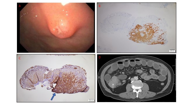
Case Description/Methods: A 59-year-old male with IgG Kappa MM with history of a left shoulder plasmacytoma (on apixaban) presented with two weeks of melena, epigastric pain and worsening anemia with a hemoglobin of 6.6 while taking NSAIDs. EGD showed four discrete atypical cratered gastric ulcers with heaped up edges and lacy vasculature in the gastric fundus and body, felt to be the source of UGIB (Fig.1a). Biopsies showed gastric mucosa with an atypical infiltrate of kappa restricted (Fig.1b), CD138 (Fig.1c), CD45 positive plasma cells, consistent with EMM. Congo red staining was negative for amyloid deposition. Bleeding resolved with continued chemotherapy and Omeprazole but he was readmitted 1 month later for a large bowel obstruction from a cecal mass requiring ileocolonic resection (Fig. 1d). Pathology of the cecal mass again revealed kappa restricted, CD138 positive plasma cells with lymphovascular invasion. PET scan confirmed rapid progression of disease. Attempts at escalating chemotherapy were made, however he developed worsening obstructive bowel symptoms and abdominal pain and was found to have extensive bowel necrosis and perforation on repeat CT imaging. After further goals of care discussion, he was switched to comfort measures and quickly died.
Discussion: EMM is seen in up to 15-30% of MM cases. However the GI tract is rarely involved, accounting for < 5% of cases. When present, it confers a poor prognosis and is a hallmark of aggressive disease. This case demonstrated two unique presentations of EMM of the GI tract - first as gastric ulcers resulting in an UGIB and subsequently as a cecal mass leading to bowel obstruction and perforation. Despite its rarity, physicians should be aware of these potential manifestations and its significance of advanced disease.

Disclosures:
Daniel Holten, MD, David Jonason, MD, Drew Ferguson, MD, Kelsey Baron, MD, Judith Staub, MD. P0775 - A Rare Case of Multiple Myeloma Manifesting as an Upper Gastrointestinal Bleed and Obstructing Cecal Mass, ACG 2024 Annual Scientific Meeting Abstracts. Philadelphia, PA: American College of Gastroenterology.
University of Utah, Salt Lake City, UT
Introduction: Multiple myeloma (MM) is a hematologic malignancy characterized by clonal proliferation of plasma cells originating in the bone marrow. It typically presents with myeloma-defining events such as hypercalcemia, renal failure, anemia and osteolytic bone lesions. Extramedullary MM (EMM) occurs when malignant plasma cells form tumors outside the bone marrow, and is associated with poor outcomes. We present a rare case of EMM with involvement of the GI tract manifesting as an acute upper gastrointestinal bleed (UGIB) and later a large bowel obstruction.
Case Description/Methods: A 59-year-old male with IgG Kappa MM with history of a left shoulder plasmacytoma (on apixaban) presented with two weeks of melena, epigastric pain and worsening anemia with a hemoglobin of 6.6 while taking NSAIDs. EGD showed four discrete atypical cratered gastric ulcers with heaped up edges and lacy vasculature in the gastric fundus and body, felt to be the source of UGIB (Fig.1a). Biopsies showed gastric mucosa with an atypical infiltrate of kappa restricted (Fig.1b), CD138 (Fig.1c), CD45 positive plasma cells, consistent with EMM. Congo red staining was negative for amyloid deposition. Bleeding resolved with continued chemotherapy and Omeprazole but he was readmitted 1 month later for a large bowel obstruction from a cecal mass requiring ileocolonic resection (Fig. 1d). Pathology of the cecal mass again revealed kappa restricted, CD138 positive plasma cells with lymphovascular invasion. PET scan confirmed rapid progression of disease. Attempts at escalating chemotherapy were made, however he developed worsening obstructive bowel symptoms and abdominal pain and was found to have extensive bowel necrosis and perforation on repeat CT imaging. After further goals of care discussion, he was switched to comfort measures and quickly died.
Discussion: EMM is seen in up to 15-30% of MM cases. However the GI tract is rarely involved, accounting for < 5% of cases. When present, it confers a poor prognosis and is a hallmark of aggressive disease. This case demonstrated two unique presentations of EMM of the GI tract - first as gastric ulcers resulting in an UGIB and subsequently as a cecal mass leading to bowel obstruction and perforation. Despite its rarity, physicians should be aware of these potential manifestations and its significance of advanced disease.

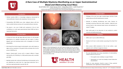
Figure: A) Non-bleeding gastric ulcer with heaped up edges and lacy vasculature, located in gastric body; B) Stomach biopsy with in-situ hybridization (ISH) for Kappa-restricted light chain plasma cell neoplasm; C) IHC stain of stomach biopsy for CD138 demonstrating a moderately well-circumscribed plasma cell neoplasm (blue arrow); D) CT A/P showing 6.9 x 3.5 cm soft tissue mass at the cecum resulting in early bowel obstruction (white arrow).
Disclosures:
Daniel Holten indicated no relevant financial relationships.
David Jonason indicated no relevant financial relationships.
Drew Ferguson indicated no relevant financial relationships.
Kelsey Baron indicated no relevant financial relationships.
Judith Staub indicated no relevant financial relationships.
Daniel Holten, MD, David Jonason, MD, Drew Ferguson, MD, Kelsey Baron, MD, Judith Staub, MD. P0775 - A Rare Case of Multiple Myeloma Manifesting as an Upper Gastrointestinal Bleed and Obstructing Cecal Mass, ACG 2024 Annual Scientific Meeting Abstracts. Philadelphia, PA: American College of Gastroenterology.
