Sunday Poster Session
Category: GI Bleeding
P0804 - Large Pedunculated Brunner Gland Hamartoma in the Duodenum Causing Massive Upper GI Bleed
Sunday, October 27, 2024
3:30 PM - 7:00 PM ET
Location: Exhibit Hall E

Has Audio
- HJ
Hasan Jaber, MD
University of Kansas School of Medicine
Wichita, KS
Presenting Author(s)
Hasan Jaber, MD, Mahmoud Mahdi, MD, Nader Al Souky, MD, Daly Al-Hadeethi, MD, Nathan Tofteland, MD, William J. Salyers, MD, MPH
University of Kansas School of Medicine, Wichita, KS
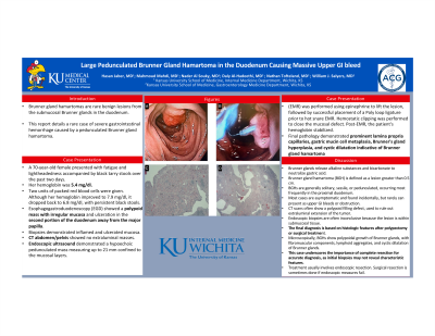
Introduction: Brunner gland hamartomas are rare benign lesions from the submucosal Brunner glands in the duodenum. This report details a rare case of severe gastrointestinal hemorrhage caused by a pedunculated Brunner gland hamartoma.
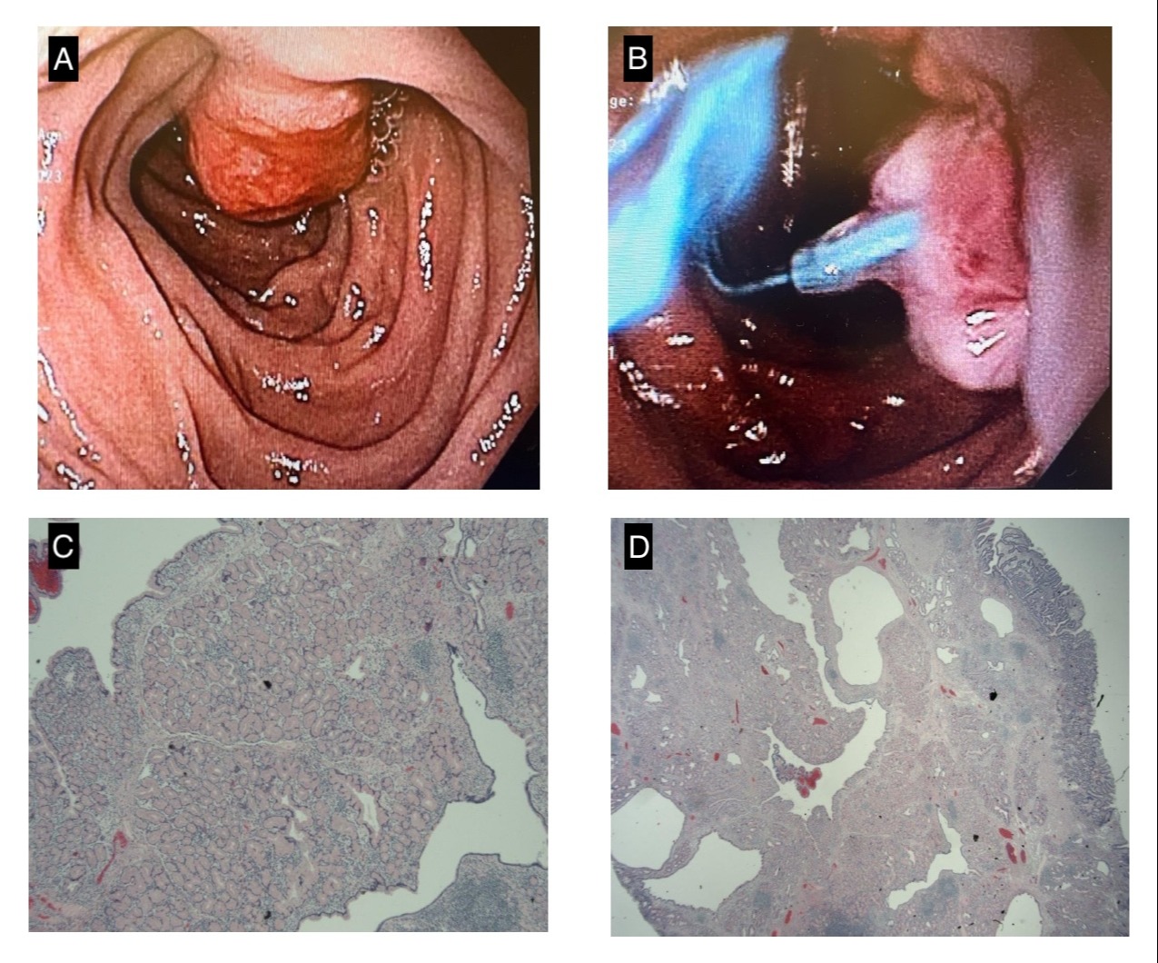
Case Description/Methods: A 70-year-old female presented with fatigue and lightheadedness accompanied by black tarry stools over the past two days. Her hemoglobin was 5.4 mg/dl. Two units of packed red blood cells were given. Although her hemoglobin improved to 7.9 mg/dl, it dropped back to 6.8 mg/dl, with persistent black stools. Esophagogastroduodenoscopy (EGD) showed a polypoid mass with irregular mucosa and ulceration in the second portion of the duodenum away from the major papilla. Biopsies demonstrated inflamed and ulcerated mucosa. A subsequent CT abdomen/pelvis showed no extraluminal masses. Endoscopic ultrasound demonstrated a hypoechoic pedunculated mass measuring up to 21 mm confined to the mucosal layers. Subsequent EGD for endoscopic mucosal resection (EMR) was performed using epinephrine to lift the lesion, followed by successful placement of a Poly loop ligature prior to hot snare EMR. Hemostatic clipping was performed to close the mucosal defect. Post-EMR, the patient's hemoglobin stabilized. Final pathology demonstrated prominent lamina propria capillaries, gastric mucin cell metaplasia, Brunner's gland hyperplasia, and cystic dilatation indicative of Brunner gland hamartoma
Discussion: Brunner glands release alkaline substances and bicarbonate to neutralize gastric acid. Brunner gland hamartoma (BGH) is defined as a lesion greater than 0.5 cm. BGHs are generally solitary, sessile, or pedunculated, occurring most frequently in the proximal duodenum. Most cases are asymptomatic and found incidentally, but rarely can present as upper GI bleeds or obstruction. CT scans often show a polypoid filling defect, used to rule out extraluminal extension of the tumor. Endoscopic biopsies are often inconclusive because the lesion is within submucosal tissue. The final diagnosis is based on histologic features after polypectomy or surgical treatment. Microscopically, BGHs show polypoidal growth of Brunner glands, with fibromuscular components, lymphoid aggregates, and cystic dilatation of Brunner glands. This case underscores the importance of complete resection for accurate diagnosis, as initial biopsies may not reveal characteristic features. Treatment usually involves endoscopic resection. Surgical resection is sometimes done if endoscopic measures fail.

Disclosures:
Hasan Jaber, MD, Mahmoud Mahdi, MD, Nader Al Souky, MD, Daly Al-Hadeethi, MD, Nathan Tofteland, MD, William J. Salyers, MD, MPH. P0804 - Large Pedunculated Brunner Gland Hamartoma in the Duodenum Causing Massive Upper GI Bleed, ACG 2024 Annual Scientific Meeting Abstracts. Philadelphia, PA: American College of Gastroenterology.
University of Kansas School of Medicine, Wichita, KS
Introduction: Brunner gland hamartomas are rare benign lesions from the submucosal Brunner glands in the duodenum. This report details a rare case of severe gastrointestinal hemorrhage caused by a pedunculated Brunner gland hamartoma.
Case Description/Methods: A 70-year-old female presented with fatigue and lightheadedness accompanied by black tarry stools over the past two days. Her hemoglobin was 5.4 mg/dl. Two units of packed red blood cells were given. Although her hemoglobin improved to 7.9 mg/dl, it dropped back to 6.8 mg/dl, with persistent black stools. Esophagogastroduodenoscopy (EGD) showed a polypoid mass with irregular mucosa and ulceration in the second portion of the duodenum away from the major papilla. Biopsies demonstrated inflamed and ulcerated mucosa. A subsequent CT abdomen/pelvis showed no extraluminal masses. Endoscopic ultrasound demonstrated a hypoechoic pedunculated mass measuring up to 21 mm confined to the mucosal layers. Subsequent EGD for endoscopic mucosal resection (EMR) was performed using epinephrine to lift the lesion, followed by successful placement of a Poly loop ligature prior to hot snare EMR. Hemostatic clipping was performed to close the mucosal defect. Post-EMR, the patient's hemoglobin stabilized. Final pathology demonstrated prominent lamina propria capillaries, gastric mucin cell metaplasia, Brunner's gland hyperplasia, and cystic dilatation indicative of Brunner gland hamartoma
Discussion: Brunner glands release alkaline substances and bicarbonate to neutralize gastric acid. Brunner gland hamartoma (BGH) is defined as a lesion greater than 0.5 cm. BGHs are generally solitary, sessile, or pedunculated, occurring most frequently in the proximal duodenum. Most cases are asymptomatic and found incidentally, but rarely can present as upper GI bleeds or obstruction. CT scans often show a polypoid filling defect, used to rule out extraluminal extension of the tumor. Endoscopic biopsies are often inconclusive because the lesion is within submucosal tissue. The final diagnosis is based on histologic features after polypectomy or surgical treatment. Microscopically, BGHs show polypoidal growth of Brunner glands, with fibromuscular components, lymphoid aggregates, and cystic dilatation of Brunner glands. This case underscores the importance of complete resection for accurate diagnosis, as initial biopsies may not reveal characteristic features. Treatment usually involves endoscopic resection. Surgical resection is sometimes done if endoscopic measures fail.

Figure: Figure 1: A&B : Brunner Gland Hamartoma in the second portion of the dueodonum C: Low power field with H&E stain D: Medium power field with H&E stain
Disclosures:
Hasan Jaber indicated no relevant financial relationships.
Mahmoud Mahdi indicated no relevant financial relationships.
Nader Al Souky indicated no relevant financial relationships.
Daly Al-Hadeethi indicated no relevant financial relationships.
Nathan Tofteland indicated no relevant financial relationships.
William Salyers indicated no relevant financial relationships.
Hasan Jaber, MD, Mahmoud Mahdi, MD, Nader Al Souky, MD, Daly Al-Hadeethi, MD, Nathan Tofteland, MD, William J. Salyers, MD, MPH. P0804 - Large Pedunculated Brunner Gland Hamartoma in the Duodenum Causing Massive Upper GI Bleed, ACG 2024 Annual Scientific Meeting Abstracts. Philadelphia, PA: American College of Gastroenterology.
