Sunday Poster Session
Category: GI Bleeding
P0810 - A New Source of Upper GI Bleeding in a Patient With Gastrostomy Tube: A Case Report
Sunday, October 27, 2024
3:30 PM - 7:00 PM ET
Location: Exhibit Hall E

Has Audio
- TH
Taraneh Honarparvar, MD
Nova Southeastern University
Wellington, FL
Presenting Author(s)
Taraneh Honarparvar, MD1, Admir Syla, MD2, Brian Hudes, MD3
1Nova Southeastern University, Wellington, FL; 2Wellington Regional Medical Center, Loxahatchee, FL; 3Wellington Regional Medical Center, Cumming, GA
Introduction:
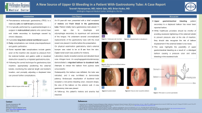
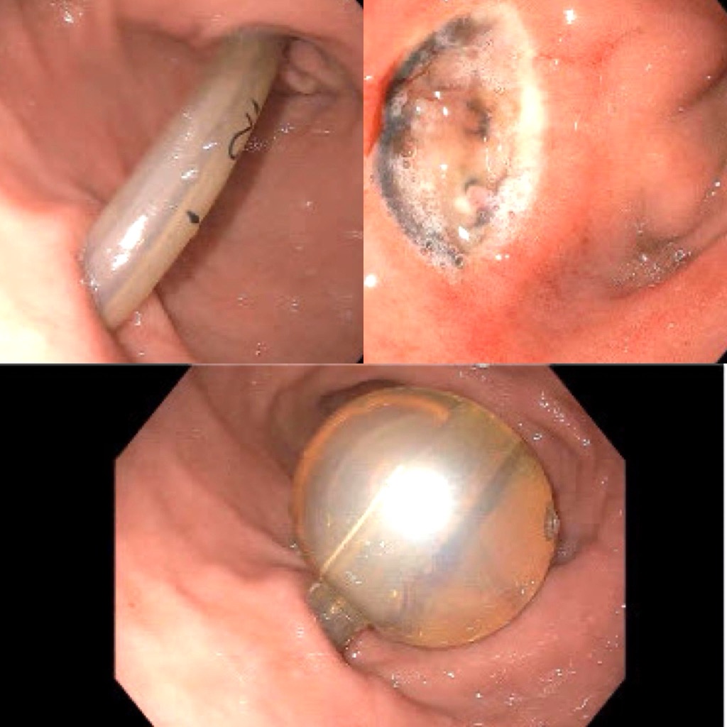
Case Description/Methods: We present a case of an elderly man who presented with a chief complaint of melena and fresh blood in the gastrostomy tube. Patient initially had a gastrostomy tube placed 11 years ago secondary to dysphagia and odynophagia resulting from squamous cell carcinoma of the tongue. He underwent several uncomplicated replacements of his gastrostomy tube with the most recent one placed 4 months before presentation. On physical examination gastrostomy tube's external bumper was noted to be at 8 cm. Digital rectal exam revealed black stool. The laboratory results revealed anemia and elevated blood urea nitrogen level. An esophagogastroduodenoscopy revealed a migrated balloon to duodenal bulb (Top left image). Attempts to retract the balloon from the pylorus were unsuccessful. Subsequently it was deflated, the tube was retracted and it was inflated to demonstrate patency (Bottom image). Endoscopic visualization of duodenal bulb revealed a non-bleeding ulcer crescent shaped ulcer the size of the balloon on the anterior wall (Top right image). A new gastrostomy tube was placed. At follow-up, the patient's melena and anemia had resolved.
Discussion: Upper gastrointestinal bleeding secondary to a displaced balloon has never been reported before. While healthcare providers should be mindful of avoiding excessive tightening of the external bolster to prevent pressure ulcers at the site of insertion, they should also recognize the risk of displacement if the external bolster is too loose. This case highlights the possibility of upper gastrointestinal bleeding secondary to a dislodged balloon causing a pressure ulcer and active bleeding in the duodenal bulb.

Disclosures:
Taraneh Honarparvar, MD1, Admir Syla, MD2, Brian Hudes, MD3. P0810 - A New Source of Upper GI Bleeding in a Patient With Gastrostomy Tube: A Case Report, ACG 2024 Annual Scientific Meeting Abstracts. Philadelphia, PA: American College of Gastroenterology.
1Nova Southeastern University, Wellington, FL; 2Wellington Regional Medical Center, Loxahatchee, FL; 3Wellington Regional Medical Center, Cumming, GA
Introduction:
Case Description/Methods: We present a case of an elderly man who presented with a chief complaint of melena and fresh blood in the gastrostomy tube. Patient initially had a gastrostomy tube placed 11 years ago secondary to dysphagia and odynophagia resulting from squamous cell carcinoma of the tongue. He underwent several uncomplicated replacements of his gastrostomy tube with the most recent one placed 4 months before presentation. On physical examination gastrostomy tube's external bumper was noted to be at 8 cm. Digital rectal exam revealed black stool. The laboratory results revealed anemia and elevated blood urea nitrogen level. An esophagogastroduodenoscopy revealed a migrated balloon to duodenal bulb (Top left image). Attempts to retract the balloon from the pylorus were unsuccessful. Subsequently it was deflated, the tube was retracted and it was inflated to demonstrate patency (Bottom image). Endoscopic visualization of duodenal bulb revealed a non-bleeding ulcer crescent shaped ulcer the size of the balloon on the anterior wall (Top right image). A new gastrostomy tube was placed. At follow-up, the patient's melena and anemia had resolved.
Discussion: Upper gastrointestinal bleeding secondary to a displaced balloon has never been reported before. While healthcare providers should be mindful of avoiding excessive tightening of the external bolster to prevent pressure ulcers at the site of insertion, they should also recognize the risk of displacement if the external bolster is too loose. This case highlights the possibility of upper gastrointestinal bleeding secondary to a dislodged balloon causing a pressure ulcer and active bleeding in the duodenal bulb.

Figure: Top left image: displaced PEG tube balloon outside the stomach. Top right image: crescent-shaped ulcer with active bleeding in the duodenal bulb. Bottom image: confirmed repositioned balloon and its patency after reinflation in the stomach.
Disclosures:
Taraneh Honarparvar indicated no relevant financial relationships.
Admir Syla indicated no relevant financial relationships.
Brian Hudes indicated no relevant financial relationships.
Taraneh Honarparvar, MD1, Admir Syla, MD2, Brian Hudes, MD3. P0810 - A New Source of Upper GI Bleeding in a Patient With Gastrostomy Tube: A Case Report, ACG 2024 Annual Scientific Meeting Abstracts. Philadelphia, PA: American College of Gastroenterology.
