Sunday Poster Session
Category: IBD
P0988 - Livedoid Vasculopathy: A Rare Cutaneous Extraintestinal Manifestation of Inflammatory Bowel Disease
Sunday, October 27, 2024
3:30 PM - 7:00 PM ET
Location: Exhibit Hall E

Has Audio
- JH
Jake Herbert, DO, MS
Oregon Health & Science University
Portland, OR
Presenting Author(s)
Jake Herbert, DO, MS, Victoria E.. Orfaly, MD, David Harmon, MD, MPH, Alex G. Ortega Loayza, MD, MCR
Oregon Health & Science University, Portland, OR
Introduction: Livedoid vasculopathy (LV) is a rare cutaneous thrombotic vasculopathy (CTV) characterized by painful relapsing ulcers. The incidence of LV is estimated to be 1:100,000, primary affects women, and is associated with rheumatologic disorders and inflammatory bowel disease (IBD). Due to its uncommonness, lack of provider familiarity, and low specificity of skin biopsy, the diagnosis of LV is often delayed. We present a case of a young woman with Crohn’s disease who presented with left ankle swelling and painful skin ulcers.
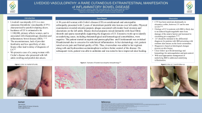
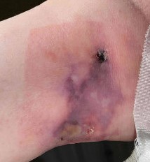
Case Description/Methods: A 38-year-old woman with Crohn’s disease on Ustekinumab and enteropathic arthropathy presented with 2 years of intermittent painful skin lesions over left ankle. Physical examination revealed reticular purpuric plaque associated with tender focal erosions and ulcerations on the left ankle. Biopsy showed purpuric mixed dermatitis with focal fibrin thrombi and sparse neutrophils supporting the diagnosis of LV. Extensive work-up to identify an underlying cause, including rheumatological and hematological comorbidities, were negative. The patient started on aspirin and pentoxyphylline and Ustekinumab was switched Risankizumab due to concerns for subclinical inflammation. At her dermatology visit, patient noted severe pain and limited quality of life. Thus, she was transitioned to rivaroxaban and duloxetine to achieve better control of the disease.
Discussion: While CTV has been noted pre-dominantly in ulcerative colitis, this is the first case of LV in a patient with Crohn’s disease reported in the literature. LV should be included in the differential diagnosis for patients with IBD who present with retiform skin lesions on the lower extremities. Etiology of LV in patients with IBD is likely due to an induced hypercoagulable state from damage of the enteric barrier and increased in circulating pro-coagulants, leading to increase von Willibrand factor and platelet dysfunction. Diagnosis is based on histological changes present on skin biopsy and patients should have close follow up with dermatology and hematology for anticoagulation and pain control in addition to appropriate management of underlying IBD.

Disclosures:
Jake Herbert, DO, MS, Victoria E.. Orfaly, MD, David Harmon, MD, MPH, Alex G. Ortega Loayza, MD, MCR. P0988 - Livedoid Vasculopathy: A Rare Cutaneous Extraintestinal Manifestation of Inflammatory Bowel Disease, ACG 2024 Annual Scientific Meeting Abstracts. Philadelphia, PA: American College of Gastroenterology.
Oregon Health & Science University, Portland, OR
Introduction: Livedoid vasculopathy (LV) is a rare cutaneous thrombotic vasculopathy (CTV) characterized by painful relapsing ulcers. The incidence of LV is estimated to be 1:100,000, primary affects women, and is associated with rheumatologic disorders and inflammatory bowel disease (IBD). Due to its uncommonness, lack of provider familiarity, and low specificity of skin biopsy, the diagnosis of LV is often delayed. We present a case of a young woman with Crohn’s disease who presented with left ankle swelling and painful skin ulcers.
Case Description/Methods: A 38-year-old woman with Crohn’s disease on Ustekinumab and enteropathic arthropathy presented with 2 years of intermittent painful skin lesions over left ankle. Physical examination revealed reticular purpuric plaque associated with tender focal erosions and ulcerations on the left ankle. Biopsy showed purpuric mixed dermatitis with focal fibrin thrombi and sparse neutrophils supporting the diagnosis of LV. Extensive work-up to identify an underlying cause, including rheumatological and hematological comorbidities, were negative. The patient started on aspirin and pentoxyphylline and Ustekinumab was switched Risankizumab due to concerns for subclinical inflammation. At her dermatology visit, patient noted severe pain and limited quality of life. Thus, she was transitioned to rivaroxaban and duloxetine to achieve better control of the disease.
Discussion: While CTV has been noted pre-dominantly in ulcerative colitis, this is the first case of LV in a patient with Crohn’s disease reported in the literature. LV should be included in the differential diagnosis for patients with IBD who present with retiform skin lesions on the lower extremities. Etiology of LV in patients with IBD is likely due to an induced hypercoagulable state from damage of the enteric barrier and increased in circulating pro-coagulants, leading to increase von Willibrand factor and platelet dysfunction. Diagnosis is based on histological changes present on skin biopsy and patients should have close follow up with dermatology and hematology for anticoagulation and pain control in addition to appropriate management of underlying IBD.

Figure: Left ankle with reticular purpuric plaque and focal ulcerations
Disclosures:
Jake Herbert indicated no relevant financial relationships.
Victoria Orfaly indicated no relevant financial relationships.
David Harmon indicated no relevant financial relationships.
Alex Ortega Loayza indicated no relevant financial relationships.
Jake Herbert, DO, MS, Victoria E.. Orfaly, MD, David Harmon, MD, MPH, Alex G. Ortega Loayza, MD, MCR. P0988 - Livedoid Vasculopathy: A Rare Cutaneous Extraintestinal Manifestation of Inflammatory Bowel Disease, ACG 2024 Annual Scientific Meeting Abstracts. Philadelphia, PA: American College of Gastroenterology.
