Sunday Poster Session
Category: Interventional Endoscopy
P1101 - Endoscopic Submucosal Dissection of a MALT Lymphoma.... in the Rectum!
Sunday, October 27, 2024
3:30 PM - 7:00 PM ET
Location: Exhibit Hall E

Has Audio
- AL
Andy L. Lin, MD
University of California, Irvine
Orange, CA
Presenting Author(s)
Andy L. Lin, MD1, Marie Balfour, MD1, Shoujit Banerjee, MD2, Amirali Tavangar, MD1, Brian Mendoza, MD1, Yousif Arif, BS3, Peter H. Nguyen, MD1, Jason Samarasena, MD1, Frances Dang, MD1
1University of California, Irvine, Orange, CA; 2University of California Irvine, Orange, CA; 3University of California Irvine School of Medicine, Orange, CA
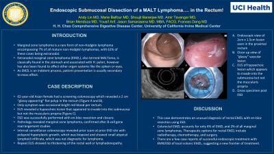
Introduction: Marginal zone lymphoma is a rare form of non-Hodgkin lymphoma encompassing 7% of all mature non-Hodgkin lymphomas, with 61% of these cases being extranodal. Extranodal marginal zone lymphoma (EMZL), also termed MALToma, is classically found in the stomach and associated with H. pylori, however has also been found to affect other organ systems like the spleen or eyes. EMZL occurs in areas of high immunologic/inflammatory activity including areas of chronic microbial infection and autoimmune disorders. As EMZL is an indolent process, patient presentation is usually secondary to mass effect. We present a case of a rectal polyp resected using endoscopic submucosal dissection (ESD) with pathology revealing EMZL.
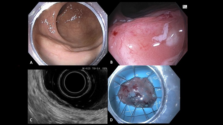
Case Description/Methods: A 62-year-old Asian female was referred to our medical center after colonoscopy revealed a 2 cm “glassy appearing” flat polyp in the rectum (Figures A and B). The patient was having occasional bright red blood per rectum. Lower endoscopic ultrasound (EUS) was performed on the 2 cm by 1.5 cm Paris IIa lesion. The lesion was hypoechoic and appeared to invade into the submucosa but not the muscularis propria (Figure C). Conventional ESD was successfully performed with en-bloc resection and closure with 5 clips. Pathology revealed marginal zone lymphoma, confirmed after B-cell rearrangement studies. There were no bulky lymphoid nodules at the periphery or deep margins. The patient returned for surveillance colonoscopy, and ESD scar was identified with polypoid hyperplastic growth, which was biopsied. Pathology of ESD scar showed small atypical lymphoid infiltrate, which was not diagnostic for lymphoma. Rectal EUS showed no thickening of the rectal wall or lymphadenopathy.
Discussion: This case demonstrates an unusual diagnosis of rectal EMZL with en-bloc resection using ESD. Colorectal EMZL accounts for only 4% of EMZL and 2% of all marginal zone lymphomas. Fortunately, EMZL has a 5-year survival rate of 94% according to recent epidemiological data. Therapeutic options for rectal EMZL include radiotherapy, chemotherapy, and surgery. There are a few case reports of successful endoscopic treatment with EMR/ESD of local colonic EMZL, suggesting a new frontier of treatment.

Disclosures:
Andy L. Lin, MD1, Marie Balfour, MD1, Shoujit Banerjee, MD2, Amirali Tavangar, MD1, Brian Mendoza, MD1, Yousif Arif, BS3, Peter H. Nguyen, MD1, Jason Samarasena, MD1, Frances Dang, MD1. P1101 - Endoscopic Submucosal Dissection of a MALT Lymphoma.... in the Rectum!, ACG 2024 Annual Scientific Meeting Abstracts. Philadelphia, PA: American College of Gastroenterology.
1University of California, Irvine, Orange, CA; 2University of California Irvine, Orange, CA; 3University of California Irvine School of Medicine, Orange, CA
Introduction: Marginal zone lymphoma is a rare form of non-Hodgkin lymphoma encompassing 7% of all mature non-Hodgkin lymphomas, with 61% of these cases being extranodal. Extranodal marginal zone lymphoma (EMZL), also termed MALToma, is classically found in the stomach and associated with H. pylori, however has also been found to affect other organ systems like the spleen or eyes. EMZL occurs in areas of high immunologic/inflammatory activity including areas of chronic microbial infection and autoimmune disorders. As EMZL is an indolent process, patient presentation is usually secondary to mass effect. We present a case of a rectal polyp resected using endoscopic submucosal dissection (ESD) with pathology revealing EMZL.
Case Description/Methods: A 62-year-old Asian female was referred to our medical center after colonoscopy revealed a 2 cm “glassy appearing” flat polyp in the rectum (Figures A and B). The patient was having occasional bright red blood per rectum. Lower endoscopic ultrasound (EUS) was performed on the 2 cm by 1.5 cm Paris IIa lesion. The lesion was hypoechoic and appeared to invade into the submucosa but not the muscularis propria (Figure C). Conventional ESD was successfully performed with en-bloc resection and closure with 5 clips. Pathology revealed marginal zone lymphoma, confirmed after B-cell rearrangement studies. There were no bulky lymphoid nodules at the periphery or deep margins. The patient returned for surveillance colonoscopy, and ESD scar was identified with polypoid hyperplastic growth, which was biopsied. Pathology of ESD scar showed small atypical lymphoid infiltrate, which was not diagnostic for lymphoma. Rectal EUS showed no thickening of the rectal wall or lymphadenopathy.
Discussion: This case demonstrates an unusual diagnosis of rectal EMZL with en-bloc resection using ESD. Colorectal EMZL accounts for only 4% of EMZL and 2% of all marginal zone lymphomas. Fortunately, EMZL has a 5-year survival rate of 94% according to recent epidemiological data. Therapeutic options for rectal EMZL include radiotherapy, chemotherapy, and surgery. There are a few case reports of successful endoscopic treatment with EMR/ESD of local colonic EMZL, suggesting a new frontier of treatment.

Figure: A. Endoscopic view of 2cm x 1.5cm lesion seen in the proximal rectum
B. Close up view of “glassy” vascular lesion
C. EUS of hypoechoic lesion which appears to invade into the submucosa but not the muscularis propria
D. Gross specimen post ESD
B. Close up view of “glassy” vascular lesion
C. EUS of hypoechoic lesion which appears to invade into the submucosa but not the muscularis propria
D. Gross specimen post ESD
Disclosures:
Andy Lin indicated no relevant financial relationships.
Marie Balfour indicated no relevant financial relationships.
Shoujit Banerjee indicated no relevant financial relationships.
Amirali Tavangar indicated no relevant financial relationships.
Brian Mendoza indicated no relevant financial relationships.
Yousif Arif indicated no relevant financial relationships.
Peter Nguyen indicated no relevant financial relationships.
Jason Samarasena: Cook Medical – Consultant. Medtronic – Advisory Committee/Board Member, Consultant. Neptune Medical – Advisory Committee/Board Member, Consultant. Olympus – Advisory Committee/Board Member, Consultant. Ovesco – Advisory Committee/Board Member, Consultant, Speakers Bureau. SatisfAI – Stock-privately held company. Steris – Advisory Committee/Board Member.
Frances Dang indicated no relevant financial relationships.
Andy L. Lin, MD1, Marie Balfour, MD1, Shoujit Banerjee, MD2, Amirali Tavangar, MD1, Brian Mendoza, MD1, Yousif Arif, BS3, Peter H. Nguyen, MD1, Jason Samarasena, MD1, Frances Dang, MD1. P1101 - Endoscopic Submucosal Dissection of a MALT Lymphoma.... in the Rectum!, ACG 2024 Annual Scientific Meeting Abstracts. Philadelphia, PA: American College of Gastroenterology.
