Sunday Poster Session
Category: Interventional Endoscopy
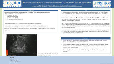
P1105 - Endoscopic Ultrasound to Diagnose Non-Neoplastic EBV-Associated Follicular Hyperplasia
Sunday, October 27, 2024
3:30 PM - 7:00 PM ET
Location: Exhibit Hall E

Has Audio
- PL
Phillip Leff, DO
Creighton University School of Medicine
Scottsdale, AZ
Presenting Author(s)
Phillip Leff, DO1, Aida Rezaie, MD2, Silpa Choday, MD3, Savio Reddymasu, MBBS, FACG4
1Creighton University School of Medicine, Scottsdale, AZ; 2Creighton University, Phoenix, AZ; 3Creighton University School of Medicine, Phoenix, AZ; 4Creighton University Medical Center, Phoenix, AZ
Introduction: Periportal lymphadenopathy on imaging studies is associated with a variety of etiologies including lymphoproliferative disorders, metastatic neoplasm, granulomatous diseases and various infections including Epstein-barr virus (EBV). EBV is also associated with a wide range of B-cell lymphoproliferative disorders. We present a rare case of periportal lymphadenopathy due to EBV in a seronegative patient determined by endoscopic ultrasound (EUS) guided lymph node biopsy.
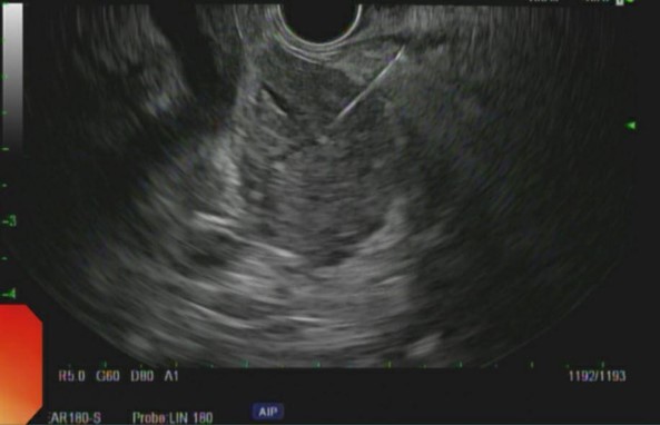
Case Description/Methods: A 31-year-old female presented with cervical lymphadenopathy, low-grade fever and a rash for several weeks. Computed tomography of the neck showed mild, bilateral lymphadenopathy up to 2 cm by 1.5 cm. She had a presumed diagnosis of non-Hodgkin's lymphoma and underwent a PET scan which revealed FDG avid lymph nodes noted in the neck, chest, abdomen and pelvis specifically surrounding the liver suggestive of non-Hodgkin's lymphoma. EUS revealed multiple enlarged periportal lymph nodes up to 3 cm amenable to fine-needle biopsy (FNB) [figure 1]. Pathology was consistent with lymphoid tissue with florid lymphoid hyperplasia and increased number of EBV positive CD30 positive cells. However, the findings were not diagnostic of a lymphoma per the WHO HAEM classification. Excisional lymph node biopsy by ENT of the neck also revealed EBV associated follicular hyperplasia. EBV PCR from the peripheral blood was however undetectable. Patient's symptoms spontaneously improved in a few weeks with conservative management.
Discussion: This is a rare case of non-neoplastic, diffuse lymphadenopathy from EBV diagnosed successfully through EUS guided FNB of the periportal lymph nodes. EUS guided FNB is the least invasive and highly effective diagnostic modality to obtain core samples from this area in comparison to traditional sampling techniques such as CT guided biopsy and excisional biopsy by laparoscopy. This case highlights the expanding role of EUS in the diagnostic algorithm of a variety of disease processes.

Disclosures:
Phillip Leff, DO1, Aida Rezaie, MD2, Silpa Choday, MD3, Savio Reddymasu, MBBS, FACG4. P1105 - Endoscopic Ultrasound to Diagnose Non-Neoplastic EBV-Associated Follicular Hyperplasia, ACG 2024 Annual Scientific Meeting Abstracts. Philadelphia, PA: American College of Gastroenterology.
1Creighton University School of Medicine, Scottsdale, AZ; 2Creighton University, Phoenix, AZ; 3Creighton University School of Medicine, Phoenix, AZ; 4Creighton University Medical Center, Phoenix, AZ
Introduction: Periportal lymphadenopathy on imaging studies is associated with a variety of etiologies including lymphoproliferative disorders, metastatic neoplasm, granulomatous diseases and various infections including Epstein-barr virus (EBV). EBV is also associated with a wide range of B-cell lymphoproliferative disorders. We present a rare case of periportal lymphadenopathy due to EBV in a seronegative patient determined by endoscopic ultrasound (EUS) guided lymph node biopsy.
Case Description/Methods: A 31-year-old female presented with cervical lymphadenopathy, low-grade fever and a rash for several weeks. Computed tomography of the neck showed mild, bilateral lymphadenopathy up to 2 cm by 1.5 cm. She had a presumed diagnosis of non-Hodgkin's lymphoma and underwent a PET scan which revealed FDG avid lymph nodes noted in the neck, chest, abdomen and pelvis specifically surrounding the liver suggestive of non-Hodgkin's lymphoma. EUS revealed multiple enlarged periportal lymph nodes up to 3 cm amenable to fine-needle biopsy (FNB) [figure 1]. Pathology was consistent with lymphoid tissue with florid lymphoid hyperplasia and increased number of EBV positive CD30 positive cells. However, the findings were not diagnostic of a lymphoma per the WHO HAEM classification. Excisional lymph node biopsy by ENT of the neck also revealed EBV associated follicular hyperplasia. EBV PCR from the peripheral blood was however undetectable. Patient's symptoms spontaneously improved in a few weeks with conservative management.
Discussion: This is a rare case of non-neoplastic, diffuse lymphadenopathy from EBV diagnosed successfully through EUS guided FNB of the periportal lymph nodes. EUS guided FNB is the least invasive and highly effective diagnostic modality to obtain core samples from this area in comparison to traditional sampling techniques such as CT guided biopsy and excisional biopsy by laparoscopy. This case highlights the expanding role of EUS in the diagnostic algorithm of a variety of disease processes.

Figure: Endoscopic ultrasound of 3-centimeter periportal lymph node
Disclosures:
Phillip Leff indicated no relevant financial relationships.
Aida Rezaie indicated no relevant financial relationships.
Silpa Choday indicated no relevant financial relationships.
Savio Reddymasu indicated no relevant financial relationships.
Phillip Leff, DO1, Aida Rezaie, MD2, Silpa Choday, MD3, Savio Reddymasu, MBBS, FACG4. P1105 - Endoscopic Ultrasound to Diagnose Non-Neoplastic EBV-Associated Follicular Hyperplasia, ACG 2024 Annual Scientific Meeting Abstracts. Philadelphia, PA: American College of Gastroenterology.
