Sunday Poster Session
Category: Interventional Endoscopy
P1109 - A Staged Approach to Resecting a Large Rectal Polyp Using Endoscopic Mucosal Resection and Trans-Anal Endoscopic Microsurgery
Sunday, October 27, 2024
3:30 PM - 7:00 PM ET
Location: Exhibit Hall E

Has Audio
- BG
Bhavna Guduguntla, MD
Washington University School of Medicine in St. Louis
St. Louis, MO
Presenting Author(s)
Bhavna Guduguntla, MD1, Jared Ye, MS1, Paul Wise, MD1, Ahmad Najdat Bazarbashi, MD2
1Washington University School of Medicine in St. Louis, St. Louis, MO; 2Washington University School of Medicine in St. Louis / Barnes-Jewish Hospital, St. Louis, MO
Introduction: Endoscopic mucosal resection (EMR) and endoscopic submucosal dissection (ESD) are first-line interventions for rectal polyps. When lesions are not amenable to either of these, surgical intervention may be necessary. Trans-anal endoscopic microsurgery (TEMS) is a less invasive, organ sparing intervention for polyp resection, however not all lesions are amenable to TEMS. We present a case of a 62-year-old woman with a 60 mm rectal polyp, resected using a staged approach with EMR followed by TEMS.
Case Description/Methods: A 62 year old female presented with 8 years of watery diarrhea. Physical exam and laboratory workup were unremarkable. She had not had prior endoscopic evaluation. Colonoscopy revealed a large 60 mm Paris Is rectal polyp [Figure 1A]. Initial biopsies confirmed tubulovillous adenoma. ESD was not thought to be amenable given location and the lesion was too large for TEMS. Therefore, EMR was pursued which was successful in removing 70% of the lesion. However, a central scarred area raised concerns of deeper submucosal involvement, preventing complete resection [Figure 1B]. This measured 25mm in diameter and could not be resected [Figure 1C]. The patient was recommended to undergo TEMS to remove residual polyp. A pelvis MRI revealed no evidence of invasion into submucosal or muscularis layer. At 3 months follow up, TEMS was performed to excise the remaining 25mm en-bloc. Pathology confirmed complete excision with negative deep and lateral margins. There was no evidence of high grade dysplasia or malignancy. The patient experienced resolution of symptoms, and at six-month follow-up sigmoidoscopy revealed a well healed scar without evidence of residual or recurrent disease [Figure 1D].
Discussion: This case demonstrates an effective staged approach for a large rectal polyp that was not amenable to complete resection with endoscopy. This approach is a less invasive alternative to radical surgery and is organ preserving. Providers should consider a similar staged approach for non-cancerous large rectal polyps that cannot be safely and entirely removed endoscopically.

Disclosures:
Bhavna Guduguntla, MD1, Jared Ye, MS1, Paul Wise, MD1, Ahmad Najdat Bazarbashi, MD2. P1109 - A Staged Approach to Resecting a Large Rectal Polyp Using Endoscopic Mucosal Resection and Trans-Anal Endoscopic Microsurgery, ACG 2024 Annual Scientific Meeting Abstracts. Philadelphia, PA: American College of Gastroenterology.
1Washington University School of Medicine in St. Louis, St. Louis, MO; 2Washington University School of Medicine in St. Louis / Barnes-Jewish Hospital, St. Louis, MO
Introduction: Endoscopic mucosal resection (EMR) and endoscopic submucosal dissection (ESD) are first-line interventions for rectal polyps. When lesions are not amenable to either of these, surgical intervention may be necessary. Trans-anal endoscopic microsurgery (TEMS) is a less invasive, organ sparing intervention for polyp resection, however not all lesions are amenable to TEMS. We present a case of a 62-year-old woman with a 60 mm rectal polyp, resected using a staged approach with EMR followed by TEMS.
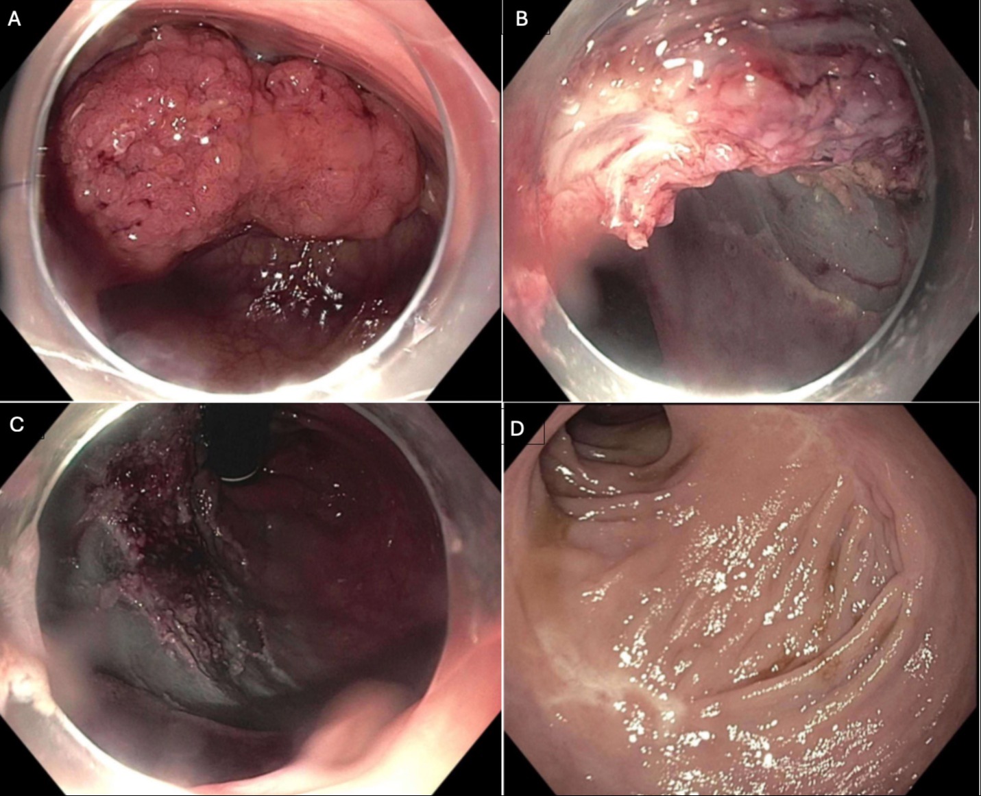
Case Description/Methods: A 62 year old female presented with 8 years of watery diarrhea. Physical exam and laboratory workup were unremarkable. She had not had prior endoscopic evaluation. Colonoscopy revealed a large 60 mm Paris Is rectal polyp [Figure 1A]. Initial biopsies confirmed tubulovillous adenoma. ESD was not thought to be amenable given location and the lesion was too large for TEMS. Therefore, EMR was pursued which was successful in removing 70% of the lesion. However, a central scarred area raised concerns of deeper submucosal involvement, preventing complete resection [Figure 1B]. This measured 25mm in diameter and could not be resected [Figure 1C]. The patient was recommended to undergo TEMS to remove residual polyp. A pelvis MRI revealed no evidence of invasion into submucosal or muscularis layer. At 3 months follow up, TEMS was performed to excise the remaining 25mm en-bloc. Pathology confirmed complete excision with negative deep and lateral margins. There was no evidence of high grade dysplasia or malignancy. The patient experienced resolution of symptoms, and at six-month follow-up sigmoidoscopy revealed a well healed scar without evidence of residual or recurrent disease [Figure 1D].
Discussion: This case demonstrates an effective staged approach for a large rectal polyp that was not amenable to complete resection with endoscopy. This approach is a less invasive alternative to radical surgery and is organ preserving. Providers should consider a similar staged approach for non-cancerous large rectal polyps that cannot be safely and entirely removed endoscopically.

Figure: Figure 1:
A: Large Paris Is rectal polyp
B: EMR with evidence of central scarring
C: Residual central 25mm of polyp that could not be resected with EMR
D: 6 month follow up of staged EMR followed by TEMS with evidence of well healed scar without recurrence
A: Large Paris Is rectal polyp
B: EMR with evidence of central scarring
C: Residual central 25mm of polyp that could not be resected with EMR
D: 6 month follow up of staged EMR followed by TEMS with evidence of well healed scar without recurrence
Disclosures:
Bhavna Guduguntla indicated no relevant financial relationships.
Jared Ye indicated no relevant financial relationships.
Paul Wise indicated no relevant financial relationships.
Ahmad Najdat Bazarbashi indicated no relevant financial relationships.
Bhavna Guduguntla, MD1, Jared Ye, MS1, Paul Wise, MD1, Ahmad Najdat Bazarbashi, MD2. P1109 - A Staged Approach to Resecting a Large Rectal Polyp Using Endoscopic Mucosal Resection and Trans-Anal Endoscopic Microsurgery, ACG 2024 Annual Scientific Meeting Abstracts. Philadelphia, PA: American College of Gastroenterology.
