Sunday Poster Session
Category: Liver
P1222 - Risk Factors, Clinical Presentation and Non-Invasive Liver Fibrosis Tests for Advanced Liver Fibrosis in Biopsy Diagnosed Metabolic Dysfunction-Associated Steatotic Liver Disease or Steatohepatitis
Sunday, October 27, 2024
3:30 PM - 7:00 PM ET
Location: Exhibit Hall E

Has Audio
- KH
Ke-Qin Hu, MD
University of California, Irvine
Orange, CA
Presenting Author(s)
Hien Lau, MD, Ke-Qin Hu, MD
University of California, Irvine, Orange, CA
Introduction: Further studies remain needed on the risk factors, natural history, and utilization of NITFs in MASLD. This study assessed the risk factors, clinical presentation, and diagnostic values of several NIFTs for advanced liver fibrosis, i.e., F3-4, in MASLD or MASH.
Methods: A single-center retrospective study on 258 pts with liver biopsy (LBx) diagnosis of MASLD or MASH and fibrosis stage, followed at the UCIMC Liver Clinic. Clinical, histological, imaging and laboratory data were collected for statistical analysis.
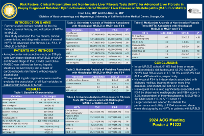
Results: The mean age was 57 ± 13; 45.6%, male; and 44.2% had diabetes mellitus (DM). All had at least one, and 61.0% had ≥ 3 risk factors; 56.4%, ≥ 3 different liver diseases (LDs); 43.2%, LBx diagnosis of MASH; 38.6%, LBx F3-4; 72.2%, FIB-4 score ≥ 1.3; 60.8% and 55.2%, ALT or AST ≥ 40 U/L; 12.2%, 20.7%, and 11.7%, albumin < 3.5 g/dL, platelets (PLTs) ≤ 120 x 109/L, or INR ≥ 1.2, respectively. On univariate analysis, LBx F3-4 (vs. F0-2) was significantly associated with DM (61.0% vs. 33.5%, p< .01), hypertension (71.0% vs. 57.6%, p=.03), bearing ≥ 3 different LDs (68.0% vs. 49.4%, p< .01) and MASH (55.0% vs. 36.1%, p< .01), but not BMI ≥ 30 (45.0% vs. 39.9%, p=.42) or hyperlipidemia (69.0% vs. 74.1%, p=.38). Additionally, LBx F3-4 was significantly associated with AST ≥ 40 U/L (64.2% vs. 50.0%, p=.04), albumin < 3.5 g/dL (20.7% vs. 7.2%, p< .01) and AFP ≥ 9 ng/mL (26.5% vs. 8.2%, p=.01). Multivariate analysis showed that DM (95% CI: 1.5-5.0; p< .01), bearing ≥ 3 LDs (95% CI: 1.3-4.7; p< .01) and MASH (95% CI: 1.2-4.1; p=.01) were significantly associated with LBx F3-4, independent of AST ≥ 40 U/L. For NIFTs, PLTs ≤ 120 x 109/L (35.4% vs. 12.3%, p< .01), F3-4 by shear wave elastography (SWE, 68.2% vs. 26.7%, p< .01), MELD-Na ≥ 10 (31.6% vs. 18.5%, p=.03), Child > 6 (24.1% vs. 7.1%, p< .01), APRI > 1 (38.0% vs. 23.4%, p=.02), and FIB-4 > 3.25 (48.1% vs. 18.2%, p< .01) were significantly associated with LBx F3-4. Multivariate analysis showed that LBx F3-4 was significantly associated with F3-4 by SWE (95% CI: 2.9-12.9; p< .01) and FIB-4 > 3.25 (95% CI: 1.1-8.6; p=.04), independent of PLTs ≤ 120 x 109/L, MELD-Na ≥ 10, Child > 6, and APRI > 1.
Discussion: In our MASLD cohort, all had at least one risk factor; 43.2%, MASH; 72.2%, FIB-4 score ≥ 1.3; 60.8% and 55.2%, ALT or AST elevation, respectively. LBx F3-4 is independently associated with DM, bearing ≥ 3 LDs, and MASH; F3-4 by SWE and FIB-4 score > 3.25, independent of thrombocytopenia, MELD-Na ≥ 10, Child > 6, or APRI score >1.
Disclosures:
Hien Lau, MD, Ke-Qin Hu, MD. P1222 - Risk Factors, Clinical Presentation and Non-Invasive Liver Fibrosis Tests for Advanced Liver Fibrosis in Biopsy Diagnosed Metabolic Dysfunction-Associated Steatotic Liver Disease or Steatohepatitis, ACG 2024 Annual Scientific Meeting Abstracts. Philadelphia, PA: American College of Gastroenterology.
University of California, Irvine, Orange, CA
Introduction: Further studies remain needed on the risk factors, natural history, and utilization of NITFs in MASLD. This study assessed the risk factors, clinical presentation, and diagnostic values of several NIFTs for advanced liver fibrosis, i.e., F3-4, in MASLD or MASH.
Methods: A single-center retrospective study on 258 pts with liver biopsy (LBx) diagnosis of MASLD or MASH and fibrosis stage, followed at the UCIMC Liver Clinic. Clinical, histological, imaging and laboratory data were collected for statistical analysis.
Results: The mean age was 57 ± 13; 45.6%, male; and 44.2% had diabetes mellitus (DM). All had at least one, and 61.0% had ≥ 3 risk factors; 56.4%, ≥ 3 different liver diseases (LDs); 43.2%, LBx diagnosis of MASH; 38.6%, LBx F3-4; 72.2%, FIB-4 score ≥ 1.3; 60.8% and 55.2%, ALT or AST ≥ 40 U/L; 12.2%, 20.7%, and 11.7%, albumin < 3.5 g/dL, platelets (PLTs) ≤ 120 x 109/L, or INR ≥ 1.2, respectively. On univariate analysis, LBx F3-4 (vs. F0-2) was significantly associated with DM (61.0% vs. 33.5%, p< .01), hypertension (71.0% vs. 57.6%, p=.03), bearing ≥ 3 different LDs (68.0% vs. 49.4%, p< .01) and MASH (55.0% vs. 36.1%, p< .01), but not BMI ≥ 30 (45.0% vs. 39.9%, p=.42) or hyperlipidemia (69.0% vs. 74.1%, p=.38). Additionally, LBx F3-4 was significantly associated with AST ≥ 40 U/L (64.2% vs. 50.0%, p=.04), albumin < 3.5 g/dL (20.7% vs. 7.2%, p< .01) and AFP ≥ 9 ng/mL (26.5% vs. 8.2%, p=.01). Multivariate analysis showed that DM (95% CI: 1.5-5.0; p< .01), bearing ≥ 3 LDs (95% CI: 1.3-4.7; p< .01) and MASH (95% CI: 1.2-4.1; p=.01) were significantly associated with LBx F3-4, independent of AST ≥ 40 U/L. For NIFTs, PLTs ≤ 120 x 109/L (35.4% vs. 12.3%, p< .01), F3-4 by shear wave elastography (SWE, 68.2% vs. 26.7%, p< .01), MELD-Na ≥ 10 (31.6% vs. 18.5%, p=.03), Child > 6 (24.1% vs. 7.1%, p< .01), APRI > 1 (38.0% vs. 23.4%, p=.02), and FIB-4 > 3.25 (48.1% vs. 18.2%, p< .01) were significantly associated with LBx F3-4. Multivariate analysis showed that LBx F3-4 was significantly associated with F3-4 by SWE (95% CI: 2.9-12.9; p< .01) and FIB-4 > 3.25 (95% CI: 1.1-8.6; p=.04), independent of PLTs ≤ 120 x 109/L, MELD-Na ≥ 10, Child > 6, and APRI > 1.
Discussion: In our MASLD cohort, all had at least one risk factor; 43.2%, MASH; 72.2%, FIB-4 score ≥ 1.3; 60.8% and 55.2%, ALT or AST elevation, respectively. LBx F3-4 is independently associated with DM, bearing ≥ 3 LDs, and MASH; F3-4 by SWE and FIB-4 score > 3.25, independent of thrombocytopenia, MELD-Na ≥ 10, Child > 6, or APRI score >1.
Disclosures:
Hien Lau indicated no relevant financial relationships.
Ke-Qin Hu: Medrigal – Speakers Bureau.
Hien Lau, MD, Ke-Qin Hu, MD. P1222 - Risk Factors, Clinical Presentation and Non-Invasive Liver Fibrosis Tests for Advanced Liver Fibrosis in Biopsy Diagnosed Metabolic Dysfunction-Associated Steatotic Liver Disease or Steatohepatitis, ACG 2024 Annual Scientific Meeting Abstracts. Philadelphia, PA: American College of Gastroenterology.
