Sunday Poster Session
Category: Liver
P1268 - Uncommon presentation of a rare disease; Hepatocellular Carcinoma in an Adult with Alagille Syndrome
Sunday, October 27, 2024
3:30 PM - 7:00 PM ET
Location: Exhibit Hall E

Has Audio

Ahmed H. abdelwahed, MD
University of Connecticut Health Center
Conneticut, CT
Presenting Author(s)
Ahmed H. Abdelwahed, MD1, Amanda Frondella, MD2, Elizabeth Richardson, DO3, Michael Einstein, MD3
1University of Connecticut Health Center, Conneticut, CT; 2University of Connecticut Health Center, Hartford, CT; 3Hartford Hospital, Hartford, CT
Introduction: Alagille syndrome is a rare autosomal dominant developmental disorder with variable clinical manifestations that include disorders in the heart, liver, eyes, and kidneys. Hepatic defects include cholestasis secondary to paucity of biliary ducts that could be seen on liver biopsy. The risk of Hepatocellular carcinoma (HCC) in patients with Alagille syndrome is not well differentiated in the literature with little known about its incidence, etiology, or screening. We hereby present a case of a 33-year-old man with Alagille syndrome who developed hepatocellular carcinoma after a history of chronic mild cholestasis. This case report shows the importance of HCC consideration in patients with Alagille syndrome.
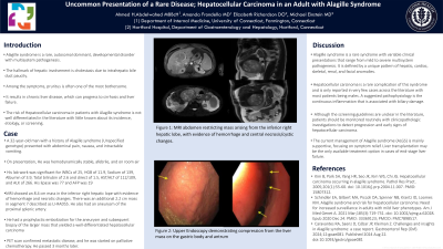
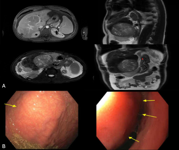
Case Description/Methods: A 32-year-old man with Alagille syndrome (Unspecified genotype) that manifested as pulmonary stenosis in childhood and chronic cholestasis presented with abdominal pain, nausea, and intractable vomiting. He did not follow up with physicians for many years. On presentation, He was hemodynamically stable. His lab work was significant for WBCs of 25, HGB of 11.9, Sodium of 139, Albumin of 3.9, Total bilirubin of 2.6 and direct of 1.5, AST/ALT of 112/109, and ALK of 266. His lipase was 77 and AFP was 19. CT abdomen and pelvis demonstrated a large irregular 8 mass in the inferior right lobe of this liver. Subsequent MRI showed an 8.4 cm mass in the inferior right hepatic lope with evidence of hemorrhage and necrotic changes. He also has an aneurysm of the proximal splenic artery measuring 2.3 x 2.3 x 2.8 cm. His liver was cirrhotic in morphology with portal hypertension that manifested by splenomegaly, portosystemic collaterals, and ascites. He had a prophylactic embolization for the aneurysm and subsequent biopsy of the larger mass that yielded a well-differentiated HCC(Grade-1). Because of his ongoing nausea and vomiting, EGD was performed and showed a deformity in the gastric body and in the gastric antrum due to extrinsic compression from a large liver mass. (Figure 2).
Discussion: HCC is a rare complication of Alagille syndrome and is only reported in very few cases with most patients being male. A suggested pathophysiology is the continuous inflammation that is associated with biliary damage. The patients should be monitored routinely with clinicopathologic investigations to detect progression and early signs of hepatocellular carcinoma. Patients might benefit from screening with abdominal ultrasound and AFP regularly frequent follow-ups for early detection of HCC.

Disclosures:
Ahmed H. Abdelwahed, MD1, Amanda Frondella, MD2, Elizabeth Richardson, DO3, Michael Einstein, MD3. P1268 - Uncommon presentation of a rare disease; Hepatocellular Carcinoma in an Adult with Alagille Syndrome, ACG 2024 Annual Scientific Meeting Abstracts. Philadelphia, PA: American College of Gastroenterology.
1University of Connecticut Health Center, Conneticut, CT; 2University of Connecticut Health Center, Hartford, CT; 3Hartford Hospital, Hartford, CT
Introduction: Alagille syndrome is a rare autosomal dominant developmental disorder with variable clinical manifestations that include disorders in the heart, liver, eyes, and kidneys. Hepatic defects include cholestasis secondary to paucity of biliary ducts that could be seen on liver biopsy. The risk of Hepatocellular carcinoma (HCC) in patients with Alagille syndrome is not well differentiated in the literature with little known about its incidence, etiology, or screening. We hereby present a case of a 33-year-old man with Alagille syndrome who developed hepatocellular carcinoma after a history of chronic mild cholestasis. This case report shows the importance of HCC consideration in patients with Alagille syndrome.
Case Description/Methods: A 32-year-old man with Alagille syndrome (Unspecified genotype) that manifested as pulmonary stenosis in childhood and chronic cholestasis presented with abdominal pain, nausea, and intractable vomiting. He did not follow up with physicians for many years. On presentation, He was hemodynamically stable. His lab work was significant for WBCs of 25, HGB of 11.9, Sodium of 139, Albumin of 3.9, Total bilirubin of 2.6 and direct of 1.5, AST/ALT of 112/109, and ALK of 266. His lipase was 77 and AFP was 19. CT abdomen and pelvis demonstrated a large irregular 8 mass in the inferior right lobe of this liver. Subsequent MRI showed an 8.4 cm mass in the inferior right hepatic lope with evidence of hemorrhage and necrotic changes. He also has an aneurysm of the proximal splenic artery measuring 2.3 x 2.3 x 2.8 cm. His liver was cirrhotic in morphology with portal hypertension that manifested by splenomegaly, portosystemic collaterals, and ascites. He had a prophylactic embolization for the aneurysm and subsequent biopsy of the larger mass that yielded a well-differentiated HCC(Grade-1). Because of his ongoing nausea and vomiting, EGD was performed and showed a deformity in the gastric body and in the gastric antrum due to extrinsic compression from a large liver mass. (Figure 2).
Discussion: HCC is a rare complication of Alagille syndrome and is only reported in very few cases with most patients being male. A suggested pathophysiology is the continuous inflammation that is associated with biliary damage. The patients should be monitored routinely with clinicopathologic investigations to detect progression and early signs of hepatocellular carcinoma. Patients might benefit from screening with abdominal ultrasound and AFP regularly frequent follow-ups for early detection of HCC.

Figure: Figure.(1)- A: MRI abdomen restricting mass arising from the inferior right
hepatic lobe, with evidence of hemorrhage and central
necrosis/cystic changes.
B:Upper Endoscopy demonstrating compression from the liver mass on the gastric body and antrum
hepatic lobe, with evidence of hemorrhage and central
necrosis/cystic changes.
B:Upper Endoscopy demonstrating compression from the liver mass on the gastric body and antrum
Disclosures:
Ahmed Abdelwahed indicated no relevant financial relationships.
Amanda Frondella indicated no relevant financial relationships.
Elizabeth Richardson indicated no relevant financial relationships.
Michael Einstein indicated no relevant financial relationships.
Ahmed H. Abdelwahed, MD1, Amanda Frondella, MD2, Elizabeth Richardson, DO3, Michael Einstein, MD3. P1268 - Uncommon presentation of a rare disease; Hepatocellular Carcinoma in an Adult with Alagille Syndrome, ACG 2024 Annual Scientific Meeting Abstracts. Philadelphia, PA: American College of Gastroenterology.
