Sunday Poster Session
Category: Liver
P1269 - Echinococcus granulosis Hydatid Liver Cyst Showing the Characteristic Water Lily Sign
Sunday, October 27, 2024
3:30 PM - 7:00 PM ET
Location: Exhibit Hall E

Has Audio

Rachael Hagen, DO
University of Connecticut Health
Farmington, CT
Presenting Author(s)
Rachael Hagen, DO1, Akaash Mittal, MD2, Neil D.. Parikh, MD3
1University of Connecticut Health, Farmington, CT; 2University of Connecticut Health Center, Farmington, CT; 3Hartford Hospital, Harford, CT
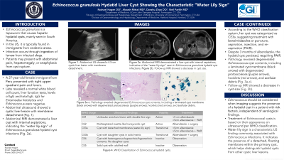
Introduction: Echinococcus granulosis is a tapeworm that causes hepatic hydatid cysts, mainly seen in South America and the Middle East. Infection occurs through ingestion of larvae from infected dogs. In America it is typically found in immigrants from endemic areas. Any organ can be affected, but hepatic involvement prevails in 65% of cases, with pulmonary involvement in 25%. Cysts are often discovered incidentally due to slow cyst growth. Patients may present with abdominal pain, hepatomegaly, or anaphylaxis from cyst rupture. It is diagnosed by abdominal US or CT scan and confirmed with serologies. We report a case of a Peruvian patient presenting with abdominal pain, whose imaging revealed a hepatic Echinococcal hydatid cyst despite negative serologies. She was successfully treated with albendazole.
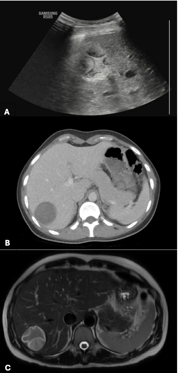
Case Description/Methods: A 27-year-old Peruvian female presented with right upper quadrant (RUQ) pain for 3 days, fevers, and a 15-kg weight loss over 3 months. Laboratory results revealed a WBC of 6.3 × 10⁹/l (eosinophils 4.1%), liver function tests were within normal limits, AFP was normal. IgE, IgG, Entamoeba histolytica IgG, and Echinococcus IgG serologies were negative. Abdominal US showed a 4.4-cm multiloculated cyst (Fig. 1a). CT abdomen showed a 4.7-cm cystic structure in the right hepatic lobe (Fig. 1b). Abdominal MRI demonstrated a 4.5-cm liver cyst with internal septations (Fig. 1c), indicative of water lily sign seen in Echinococcus infections. She was treated with oral albendazole 10 mg/kg for 3 months and monitored with abdominal US. At 1 week follow-up, she reported improvement of RUQ pain. Repeat liver function tests were normal and Echinococcus serologies remained negative.
Discussion: This case highlights the importance of maintaining a broad differential diagnosis for hepatic cysts. Echinococcus should be considered when imaging is suggestive of hydatid cysts, even in the absence of positive serologies, particularly if the characteristic water lily sign is present in a patient with risk factors. There was no pulmonary involvement on imaging, which carries a worse prognosis. Treatment of hydatid cysts is based on the WHO classification on US. Prompt identification is imperative for classification, as early intervention with benzimidazoles alone may be effective. This patient’s cyst was classified as CE3a, suggesting treatment with benzimidazoles or puncture, aspiration, injection, and re-aspiration (PAIR). PAIR was planned if the patient did not improve on anthelminthics.

Disclosures:
Rachael Hagen, DO1, Akaash Mittal, MD2, Neil D.. Parikh, MD3. P1269 - <i>Echinococcus granulosis</i> Hydatid Liver Cyst Showing the Characteristic Water Lily Sign, ACG 2024 Annual Scientific Meeting Abstracts. Philadelphia, PA: American College of Gastroenterology.
1University of Connecticut Health, Farmington, CT; 2University of Connecticut Health Center, Farmington, CT; 3Hartford Hospital, Harford, CT
Introduction: Echinococcus granulosis is a tapeworm that causes hepatic hydatid cysts, mainly seen in South America and the Middle East. Infection occurs through ingestion of larvae from infected dogs. In America it is typically found in immigrants from endemic areas. Any organ can be affected, but hepatic involvement prevails in 65% of cases, with pulmonary involvement in 25%. Cysts are often discovered incidentally due to slow cyst growth. Patients may present with abdominal pain, hepatomegaly, or anaphylaxis from cyst rupture. It is diagnosed by abdominal US or CT scan and confirmed with serologies. We report a case of a Peruvian patient presenting with abdominal pain, whose imaging revealed a hepatic Echinococcal hydatid cyst despite negative serologies. She was successfully treated with albendazole.
Case Description/Methods: A 27-year-old Peruvian female presented with right upper quadrant (RUQ) pain for 3 days, fevers, and a 15-kg weight loss over 3 months. Laboratory results revealed a WBC of 6.3 × 10⁹/l (eosinophils 4.1%), liver function tests were within normal limits, AFP was normal. IgE, IgG, Entamoeba histolytica IgG, and Echinococcus IgG serologies were negative. Abdominal US showed a 4.4-cm multiloculated cyst (Fig. 1a). CT abdomen showed a 4.7-cm cystic structure in the right hepatic lobe (Fig. 1b). Abdominal MRI demonstrated a 4.5-cm liver cyst with internal septations (Fig. 1c), indicative of water lily sign seen in Echinococcus infections. She was treated with oral albendazole 10 mg/kg for 3 months and monitored with abdominal US. At 1 week follow-up, she reported improvement of RUQ pain. Repeat liver function tests were normal and Echinococcus serologies remained negative.
Discussion: This case highlights the importance of maintaining a broad differential diagnosis for hepatic cysts. Echinococcus should be considered when imaging is suggestive of hydatid cysts, even in the absence of positive serologies, particularly if the characteristic water lily sign is present in a patient with risk factors. There was no pulmonary involvement on imaging, which carries a worse prognosis. Treatment of hydatid cysts is based on the WHO classification on US. Prompt identification is imperative for classification, as early intervention with benzimidazoles alone may be effective. This patient’s cyst was classified as CE3a, suggesting treatment with benzimidazoles or puncture, aspiration, injection, and re-aspiration (PAIR). PAIR was planned if the patient did not improve on anthelminthics.

Figure: Figure 1a. RUQ ultrasound demonstrated a complex multiloculated cystic liver lesion measuring up to 4.4 cm.
Figure 1b. CT abdomen and pelvis showed a 4.7 cm heterogenous predominantly cystic structure in the right hepatic lobe with serpiginous hyper densities.
Figure 1c. MRI demonstrated “water lily sign” in the right hepatic lobe, classically seen in Echinococcal hydatid cyst infections.
Figure 1b. CT abdomen and pelvis showed a 4.7 cm heterogenous predominantly cystic structure in the right hepatic lobe with serpiginous hyper densities.
Figure 1c. MRI demonstrated “water lily sign” in the right hepatic lobe, classically seen in Echinococcal hydatid cyst infections.
Disclosures:
Rachael Hagen indicated no relevant financial relationships.
Akaash Mittal indicated no relevant financial relationships.
Neil Parikh indicated no relevant financial relationships.
Rachael Hagen, DO1, Akaash Mittal, MD2, Neil D.. Parikh, MD3. P1269 - <i>Echinococcus granulosis</i> Hydatid Liver Cyst Showing the Characteristic Water Lily Sign, ACG 2024 Annual Scientific Meeting Abstracts. Philadelphia, PA: American College of Gastroenterology.
