Sunday Poster Session
Category: Liver
P1322 - Surgical Intervention for Symptomatic Giant Hepatic Hemangioma: A Case Report
Sunday, October 27, 2024
3:30 PM - 7:00 PM ET
Location: Exhibit Hall E

- RR
Rajmohan Rammohan, MD
Nassau University Medical Center
East Meadow, NY
Presenting Author(s)
Raj Mohan Ram Mohan, MD, Leeza Pannikodu, MD, Sai Reshma Magam, MD, Sai Greeshma Magam, MD, Melvin Joy, MD, Dilman Natt, MD, Abhishek Tadikonda, MD, Winghang Lau, MD, Jiten Desai, MD, Paul Mustacchia, MD, MBA
Nassau University Medical Center, East Meadow, NY
Introduction: Hepatic hemangiomas or hepatic venous malformations are the most common benign vascular liver lesions. They are usually asymptomatic, but when rapidly enlarging and compressing other organs, symptoms include right upper quadrant pain, early satiety, nausea and vomiting. Giant liver hemangiomas are defined by a diameter larger than 5 cm. In patients with giant liver hemangioma, observation is justified in the absence of symptoms. Surgical resection is indicated in patients with abdominal (mechanical) complaints or complications, or when diagnosis remains inconclusive.
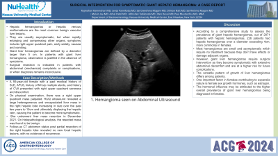
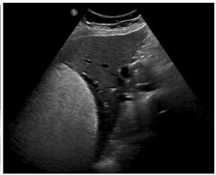
Case Description/Methods: A 59-year-old female with a past medical history of SLE, APLS, history of MI s/p multiple stents, and history of CVA presented with right upper quadrant soreness and discomfort. On physical examination, there was a right upper quadrant mass palpated. RUQ ultrasound revealed a large heterogeneous and encapsulated liver mass in the right hepatic lobe increasing in size over the past few years to 15cm and ultimately displacing the hepatic vein, causing the patient to become more symptomatic. She underwent liver mass resection in December 2021. On histopathological analysis, the resected mass was found to be benign. Follow-up CT abdomen status post partial resection of the right hepatic lobe revealed no new focal hepatic lesions, with no evidence of recurrence.
Discussion: According to a comprehensive study to assess the prevalence of giant hepatic hemangiomas, out of 2071 patients with hepatic hemangiomas, 226 patients had hepatic hemangiomas over a diameter exceeding 4cm, more commonly in females. Most hemangiomas are small and asymptomatic which require no treatment because they don’t have effects or damage adjacent organs. However, giant liver hemangiomas require surgical intervention as they become symptomatic with extensive abdominal discomfort and are at a higher risk for future complications. The versatile pattern of growth of liver hemangiomas differs among patients. One important factor in females contributing to expansile nature is female sex growth hormones, such as estrogen. The hormonal influence may be attributed to the higher overall prevalence of giant liver hemangiomas being diagnosed in females.

Disclosures:
Raj Mohan Ram Mohan, MD, Leeza Pannikodu, MD, Sai Reshma Magam, MD, Sai Greeshma Magam, MD, Melvin Joy, MD, Dilman Natt, MD, Abhishek Tadikonda, MD, Winghang Lau, MD, Jiten Desai, MD, Paul Mustacchia, MD, MBA. P1322 - Surgical Intervention for Symptomatic Giant Hepatic Hemangioma: A Case Report, ACG 2024 Annual Scientific Meeting Abstracts. Philadelphia, PA: American College of Gastroenterology.
Nassau University Medical Center, East Meadow, NY
Introduction: Hepatic hemangiomas or hepatic venous malformations are the most common benign vascular liver lesions. They are usually asymptomatic, but when rapidly enlarging and compressing other organs, symptoms include right upper quadrant pain, early satiety, nausea and vomiting. Giant liver hemangiomas are defined by a diameter larger than 5 cm. In patients with giant liver hemangioma, observation is justified in the absence of symptoms. Surgical resection is indicated in patients with abdominal (mechanical) complaints or complications, or when diagnosis remains inconclusive.
Case Description/Methods: A 59-year-old female with a past medical history of SLE, APLS, history of MI s/p multiple stents, and history of CVA presented with right upper quadrant soreness and discomfort. On physical examination, there was a right upper quadrant mass palpated. RUQ ultrasound revealed a large heterogeneous and encapsulated liver mass in the right hepatic lobe increasing in size over the past few years to 15cm and ultimately displacing the hepatic vein, causing the patient to become more symptomatic. She underwent liver mass resection in December 2021. On histopathological analysis, the resected mass was found to be benign. Follow-up CT abdomen status post partial resection of the right hepatic lobe revealed no new focal hepatic lesions, with no evidence of recurrence.
Discussion: According to a comprehensive study to assess the prevalence of giant hepatic hemangiomas, out of 2071 patients with hepatic hemangiomas, 226 patients had hepatic hemangiomas over a diameter exceeding 4cm, more commonly in females. Most hemangiomas are small and asymptomatic which require no treatment because they don’t have effects or damage adjacent organs. However, giant liver hemangiomas require surgical intervention as they become symptomatic with extensive abdominal discomfort and are at a higher risk for future complications. The versatile pattern of growth of liver hemangiomas differs among patients. One important factor in females contributing to expansile nature is female sex growth hormones, such as estrogen. The hormonal influence may be attributed to the higher overall prevalence of giant liver hemangiomas being diagnosed in females.

Figure: 1. Hemangioma seen on Abdominal Ultrasound
Disclosures:
Raj Mohan Ram Mohan indicated no relevant financial relationships.
Leeza Pannikodu indicated no relevant financial relationships.
Sai Reshma Magam indicated no relevant financial relationships.
Sai Greeshma Magam indicated no relevant financial relationships.
Melvin Joy indicated no relevant financial relationships.
Dilman Natt indicated no relevant financial relationships.
Abhishek Tadikonda indicated no relevant financial relationships.
Winghang Lau indicated no relevant financial relationships.
Jiten Desai indicated no relevant financial relationships.
Paul Mustacchia indicated no relevant financial relationships.
Raj Mohan Ram Mohan, MD, Leeza Pannikodu, MD, Sai Reshma Magam, MD, Sai Greeshma Magam, MD, Melvin Joy, MD, Dilman Natt, MD, Abhishek Tadikonda, MD, Winghang Lau, MD, Jiten Desai, MD, Paul Mustacchia, MD, MBA. P1322 - Surgical Intervention for Symptomatic Giant Hepatic Hemangioma: A Case Report, ACG 2024 Annual Scientific Meeting Abstracts. Philadelphia, PA: American College of Gastroenterology.
