Sunday Poster Session
Category: Liver
P1324 - Navigating Complexities: A Detailed Case Study of Metastatic Malignant Melanoma
Sunday, October 27, 2024
3:30 PM - 7:00 PM ET
Location: Exhibit Hall E

- AT
Abhishek Tadikonda, MD
Nassau University Medical Center
East Meadow, NY
Presenting Author(s)
Raj Mohan Ram Mohan, MD, Sai Reshma Magam, MD, Sai Greeshma Magam, MD, Melvin Joy, MD, Dilman Natt, MD, Abhishek Tadikonda, MD, Leeza Pannikodu, MD, Winghang Lau, MD, Jiten Desai, MD, Paul Mustacchia, MD, MBA
Nassau University Medical Center, East Meadow, NY
Introduction: Malignant melanoma, a cancer originating from the malignant transformation of melanocytes (neural crest cells), primarily metastasizes to lymph nodes and frequently spreads to the liver, lungs, bones, and brain. Early detection and timely surgical intervention significantly improve outcomes. The overall survival rate for those with metastatic malignant melanoma is about 5%, with typical survival ranging from 6 to 9 months. Despite surgical efforts, life expectancy usually extends to only about 12 months post-diagnosis. However, patients with isolated liver metastases exhibit a better five-year survival rate, varying significantly in literature from 10-59% to 21-22%, compared to those with widespread metastatic disease.
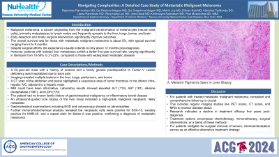
Case Description/Methods: A 52-year-old male with a history of sciatica and a family genetic predisposition to Factor V Leiden deficiency was hospitalized due to back pain. Imaging revealed multiple lesions in the liver, lungs, peritoneum, and bones. A CT scan of the abdomen and pelvis highlighted a suspicious area of tumor thrombus in the inferior infra-hepatic IVC, adjacent to a hepatic mass. MRI could have been informative. Laboratory results showed elevated ALT (110), AST (181), alkaline phosphatase (1081), and LDH (770). The patient had no known family history of gastrointestinal malignancy or inflammatory bowel disease. An ultrasound-guided core biopsy of the liver mass indicated a high-grade malignant neoplasm, likely metastatic. Gastrointestinal examinations including EGD and colonoscopy showed no abnormalities. Further immunohistochemical analysis revealed the neoplastic cells were positive for SOX-10, variably positive for HMB-45, and a repeat stain for Melan-A was positive, confirming a diagnosis of metastatic melanoma.
Discussion: For patients with treated metastatic malignant melanoma, consistent and comprehensive follow-up is crucial. This includes regular imaging studies like PET scans, CT scans, and MRIs to monitor disease status. Research indicates a decline in treatment efficacy five years post-diagnosis. Treatment options encompass chemotherapy, immunotherapy, surgical interventions, or a blend of these methods. For patients ineligible for surgical removal of tumors, chemoembolization serves as an effective alternative treatment strategy.

Disclosures:
Raj Mohan Ram Mohan, MD, Sai Reshma Magam, MD, Sai Greeshma Magam, MD, Melvin Joy, MD, Dilman Natt, MD, Abhishek Tadikonda, MD, Leeza Pannikodu, MD, Winghang Lau, MD, Jiten Desai, MD, Paul Mustacchia, MD, MBA. P1324 - Navigating Complexities: A Detailed Case Study of Metastatic Malignant Melanoma, ACG 2024 Annual Scientific Meeting Abstracts. Philadelphia, PA: American College of Gastroenterology.
Nassau University Medical Center, East Meadow, NY
Introduction: Malignant melanoma, a cancer originating from the malignant transformation of melanocytes (neural crest cells), primarily metastasizes to lymph nodes and frequently spreads to the liver, lungs, bones, and brain. Early detection and timely surgical intervention significantly improve outcomes. The overall survival rate for those with metastatic malignant melanoma is about 5%, with typical survival ranging from 6 to 9 months. Despite surgical efforts, life expectancy usually extends to only about 12 months post-diagnosis. However, patients with isolated liver metastases exhibit a better five-year survival rate, varying significantly in literature from 10-59% to 21-22%, compared to those with widespread metastatic disease.
Case Description/Methods: A 52-year-old male with a history of sciatica and a family genetic predisposition to Factor V Leiden deficiency was hospitalized due to back pain. Imaging revealed multiple lesions in the liver, lungs, peritoneum, and bones. A CT scan of the abdomen and pelvis highlighted a suspicious area of tumor thrombus in the inferior infra-hepatic IVC, adjacent to a hepatic mass. MRI could have been informative. Laboratory results showed elevated ALT (110), AST (181), alkaline phosphatase (1081), and LDH (770). The patient had no known family history of gastrointestinal malignancy or inflammatory bowel disease. An ultrasound-guided core biopsy of the liver mass indicated a high-grade malignant neoplasm, likely metastatic. Gastrointestinal examinations including EGD and colonoscopy showed no abnormalities. Further immunohistochemical analysis revealed the neoplastic cells were positive for SOX-10, variably positive for HMB-45, and a repeat stain for Melan-A was positive, confirming a diagnosis of metastatic melanoma.
Discussion: For patients with treated metastatic malignant melanoma, consistent and comprehensive follow-up is crucial. This includes regular imaging studies like PET scans, CT scans, and MRIs to monitor disease status. Research indicates a decline in treatment efficacy five years post-diagnosis. Treatment options encompass chemotherapy, immunotherapy, surgical interventions, or a blend of these methods. For patients ineligible for surgical removal of tumors, chemoembolization serves as an effective alternative treatment strategy.

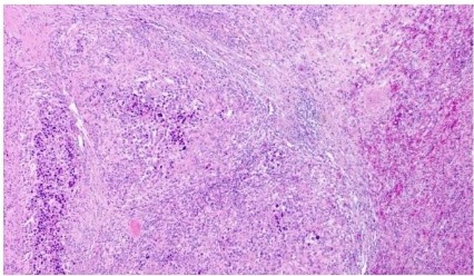
Figure: A. Melanin Pigments Seen in Liver Biopsy
Disclosures:
Raj Mohan Ram Mohan indicated no relevant financial relationships.
Sai Reshma Magam indicated no relevant financial relationships.
Sai Greeshma Magam indicated no relevant financial relationships.
Melvin Joy indicated no relevant financial relationships.
Dilman Natt indicated no relevant financial relationships.
Abhishek Tadikonda indicated no relevant financial relationships.
Leeza Pannikodu indicated no relevant financial relationships.
Winghang Lau indicated no relevant financial relationships.
Jiten Desai indicated no relevant financial relationships.
Paul Mustacchia indicated no relevant financial relationships.
Raj Mohan Ram Mohan, MD, Sai Reshma Magam, MD, Sai Greeshma Magam, MD, Melvin Joy, MD, Dilman Natt, MD, Abhishek Tadikonda, MD, Leeza Pannikodu, MD, Winghang Lau, MD, Jiten Desai, MD, Paul Mustacchia, MD, MBA. P1324 - Navigating Complexities: A Detailed Case Study of Metastatic Malignant Melanoma, ACG 2024 Annual Scientific Meeting Abstracts. Philadelphia, PA: American College of Gastroenterology.
