Sunday Poster Session
Category: Liver
P1379 - Triple Risk Factors for Budd-Chiari Syndrome
Sunday, October 27, 2024
3:30 PM - 7:00 PM ET
Location: Exhibit Hall E

Has Audio

Bianca S. Arellano, MD
University of Arizona College of Medicine
Phoenix, AZ
Presenting Author(s)
Bianca S. Arellano, MD, Elvis J. Arteaga, MD, Matthew Chaung, MD, Othniel Balizou, MD, Oscar Salinas, BS, Vanessa F. Eller, MD, Jen-Jung Pan, MD, PhD
University of Arizona College of Medicine, Phoenix, AZ
Introduction: Budd-Chiari syndrome (BCS) is a rare vascular disorder of the liver. According to the AASLD 2020 Practice Guidance on BCS, the incidence is less than 1 case per million person-years and the prevalence is 1.40-7.69 per million. While approximately 75% of patients have one underlying prothombotic condition, only 35% of patients present with more than one prothrombotic condition. The purpose of this case report is to highlight the abnormality of patient presenting with a mixture of three risk factors, and additionally finding both hepatic vein thrombosis (HVT) and portal vein thrombosis (PVT) with no history of cirrhosis.
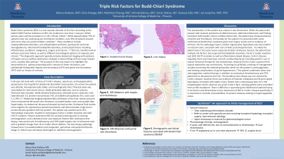
Case Description/Methods: A 36 year-old male with a history of Crohn's disease, opioid use, and hypogonadism, presented to the hospital for abdominal discomfort and jaundice. On arrival, the patient was afebrile, hemodynamically stable, and neurologically intact. Physical exam was remarkable for mild scleral icterus, mildly distended abdomen, and no asterixis. Pertinent labs included: international normalized ratio 1.7, total bilirubin 3.3, alanine transaminase 143, and alkaline phosphatase 351. Abdominal imaging indicated fatty infiltration of the liver, nearly occlusive main and proximal left portal vein thrombus, occluded hepatic veins and caudate lobe hypertrophy. An abdominal ultrasound showed minimal ascites. Peritoneal fluid studies were negative for spontaneous bacterial peritonitis and demonstrated a high serum ascites albumin gradient with low protein. The patient was worked up for BCS. Hematologic evaluation resulted in homozygous Factor V Leiden mutation and JAK2 V617F mutation. Patient underwent liver biopsy, main portal vein thrombectomy, and TIPS. The liver biopsy pathology showed zone 3 sinusoidal dilation and congestion, pericelluar and periportal fibrosis (stage 2). Patient was ultimately discharged on indefinite anticoagulation.
Discussion: The presentation of this patient was unique in two dimensions. First, the patient did not classically present with abdominal ascites or esophageal varices suggestive of portal hypertension. Given the liver biopsy results, it is likely the patient was in the acute versus subacute duration of disease. However, the patient did have both HVT and PVT, requiring medical and procedural therapies. Second, patient had multiple risk factors: two hematologic gene mutations and Crohn's disease. There is difficulty in quantifying the likelihood of patient having 3 risk factors and limited data on the mechanism of BCS in Crohn's disease.

Disclosures:
Bianca S. Arellano, MD, Elvis J. Arteaga, MD, Matthew Chaung, MD, Othniel Balizou, MD, Oscar Salinas, BS, Vanessa F. Eller, MD, Jen-Jung Pan, MD, PhD. P1379 - Triple Risk Factors for Budd-Chiari Syndrome, ACG 2024 Annual Scientific Meeting Abstracts. Philadelphia, PA: American College of Gastroenterology.
University of Arizona College of Medicine, Phoenix, AZ
Introduction: Budd-Chiari syndrome (BCS) is a rare vascular disorder of the liver. According to the AASLD 2020 Practice Guidance on BCS, the incidence is less than 1 case per million person-years and the prevalence is 1.40-7.69 per million. While approximately 75% of patients have one underlying prothombotic condition, only 35% of patients present with more than one prothrombotic condition. The purpose of this case report is to highlight the abnormality of patient presenting with a mixture of three risk factors, and additionally finding both hepatic vein thrombosis (HVT) and portal vein thrombosis (PVT) with no history of cirrhosis.
Case Description/Methods: A 36 year-old male with a history of Crohn's disease, opioid use, and hypogonadism, presented to the hospital for abdominal discomfort and jaundice. On arrival, the patient was afebrile, hemodynamically stable, and neurologically intact. Physical exam was remarkable for mild scleral icterus, mildly distended abdomen, and no asterixis. Pertinent labs included: international normalized ratio 1.7, total bilirubin 3.3, alanine transaminase 143, and alkaline phosphatase 351. Abdominal imaging indicated fatty infiltration of the liver, nearly occlusive main and proximal left portal vein thrombus, occluded hepatic veins and caudate lobe hypertrophy. An abdominal ultrasound showed minimal ascites. Peritoneal fluid studies were negative for spontaneous bacterial peritonitis and demonstrated a high serum ascites albumin gradient with low protein. The patient was worked up for BCS. Hematologic evaluation resulted in homozygous Factor V Leiden mutation and JAK2 V617F mutation. Patient underwent liver biopsy, main portal vein thrombectomy, and TIPS. The liver biopsy pathology showed zone 3 sinusoidal dilation and congestion, pericelluar and periportal fibrosis (stage 2). Patient was ultimately discharged on indefinite anticoagulation.
Discussion: The presentation of this patient was unique in two dimensions. First, the patient did not classically present with abdominal ascites or esophageal varices suggestive of portal hypertension. Given the liver biopsy results, it is likely the patient was in the acute versus subacute duration of disease. However, the patient did have both HVT and PVT, requiring medical and procedural therapies. Second, patient had multiple risk factors: two hematologic gene mutations and Crohn's disease. There is difficulty in quantifying the likelihood of patient having 3 risk factors and limited data on the mechanism of BCS in Crohn's disease.

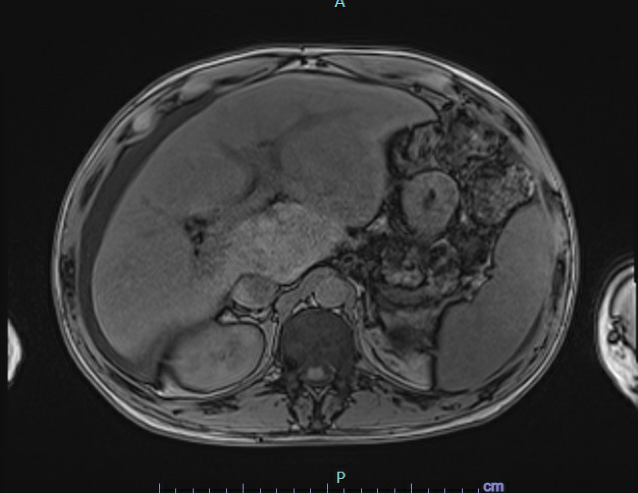
Figure: MRI Abdomen/Pelvis with and without contrast.
Disclosures:
Bianca Arellano indicated no relevant financial relationships.
Elvis Arteaga indicated no relevant financial relationships.
Matthew Chaung indicated no relevant financial relationships.
Othniel Balizou indicated no relevant financial relationships.
Oscar Salinas indicated no relevant financial relationships.
Vanessa Eller indicated no relevant financial relationships.
Jen-Jung Pan indicated no relevant financial relationships.
Bianca S. Arellano, MD, Elvis J. Arteaga, MD, Matthew Chaung, MD, Othniel Balizou, MD, Oscar Salinas, BS, Vanessa F. Eller, MD, Jen-Jung Pan, MD, PhD. P1379 - Triple Risk Factors for Budd-Chiari Syndrome, ACG 2024 Annual Scientific Meeting Abstracts. Philadelphia, PA: American College of Gastroenterology.
