Sunday Poster Session
Category: Liver
P1390 - Hepatocellular Injury From Cyst Compression in Polycystic Kidney Disease
Sunday, October 27, 2024
3:30 PM - 7:00 PM ET
Location: Exhibit Hall E

Has Audio

James Valderrama, MD, MS
University of Chicago Medicine
Chicago, IL
Presenting Author(s)
James Valderrama, MD, MS, Sonali Paul, MD, MSc
University of Chicago Medicine, Chicago, IL
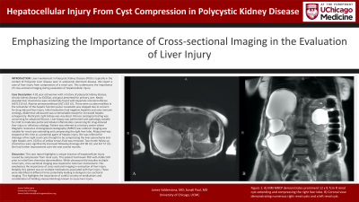
Introduction: Liver involvement in Polycystic Kidney Disease (PKD) is typically in the context of Polycystic Liver disease seen in autosomal dominant disease. We report a case of liver injury from compression of a renal cyst. This underscores the importance of cross-sectional imaging during evaluation of hepatocellular injury.
Case Description/Methods: A 82 year old woman with a history of polycystic kidney disease, chronic kidney disease 3a (CKD3a), and gout presented for primary care. Newly elevated liver chemistries were incidentally found with Aspartate aminotransferase (AST) 219 U/L Alanine aminotransferase (ALT) 333 U/L. There were no abnormalities in the remainder of the hepatic function panel. Lovastatin was stopped due to concern for drug induced liver injury. Initial evaluation had negative hepatitis and auto-immune serology.
Abdominal ultrasound was unremarkable except for increased hepatic echogenicity. Multicystic right kidney was visualized. Fibrosis serological testing was concerning for advanced fibrosis. Liver biopsy was performed with pathology notable for mild to moderate portal and lobular inflammation concerning for drug-induced liver injury or infectious etiology.
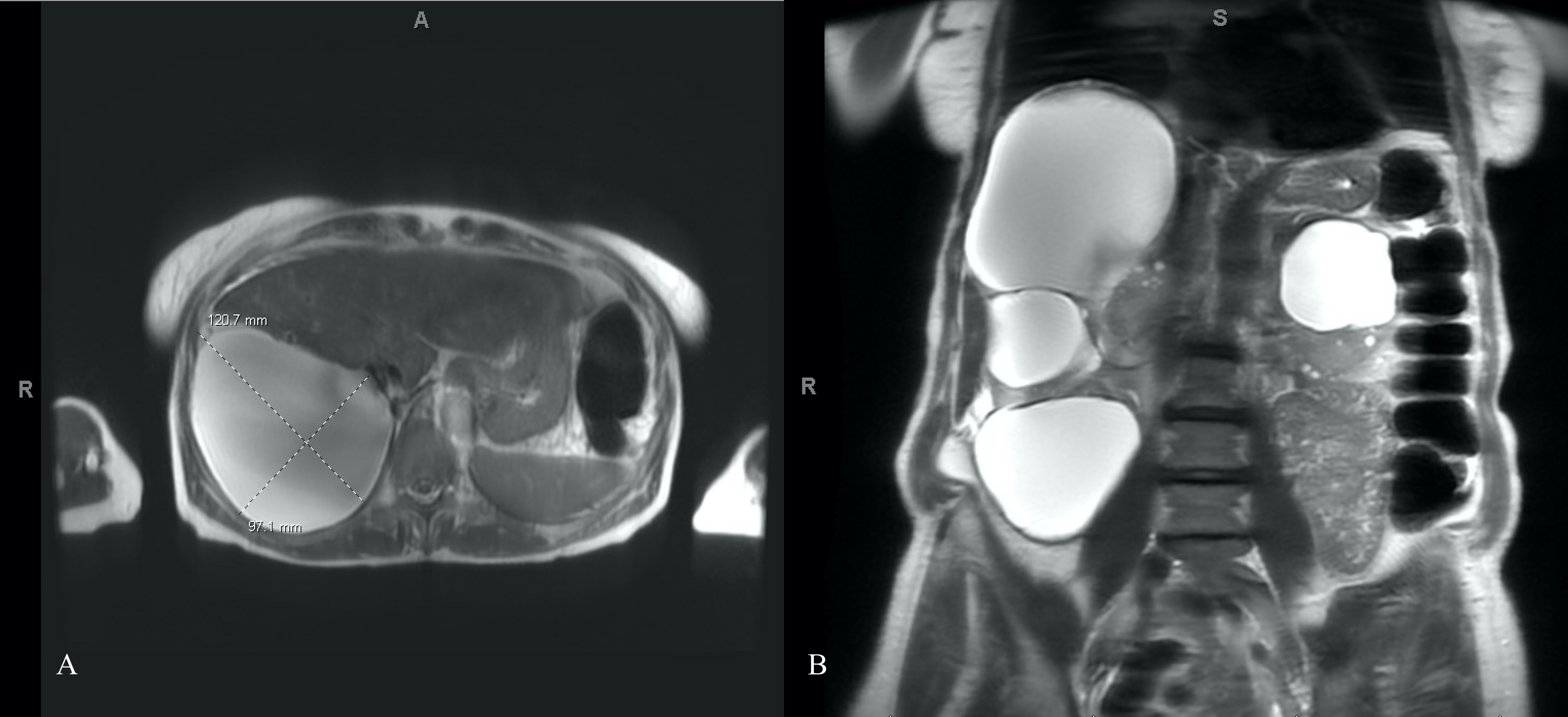
Patient was referred to a tertiary center where Magnetic resonance cholangiopancreatography (MRCP) was ordered. Imaging was notable for renal cysts extending and compressing the right liver lobe. Allopurinol was stopped at this time as a potential agent of hepatic injury.
She was referred for drainage of her right renal cysts thought to be compressing the liver parenchyma and right hepatic vein. 1020 cc of yellow serous fluid was removed. Two month follow up chemistries were significantly improved following drainage AST 90 U/L and ALT 57 U/L. She had further improvement over the next several months.
Discussion: This case report highlights a unique instance of hepatocellular injury caused by compression from renal cysts. This patient had known PKD with stable CKD prior to initial liver chemistry abnormalities. While ultrasound did visualize multiple renal cysts, cross-sectional imaging was required to note liver involvement. This emphasizes the importance of cross-sectional imaging in evaluation of liver injury.
Notably, this patient was on multiple medications associated with liver injury. These were identified at different times potentially leading to delayed cross-sectional imaging. This highlights the importance of careful scrutiny of medications and consideration of holding unessential drugs known to cause liver injury.

Disclosures:
James Valderrama, MD, MS, Sonali Paul, MD, MSc. P1390 - Hepatocellular Injury From Cyst Compression in Polycystic Kidney Disease, ACG 2024 Annual Scientific Meeting Abstracts. Philadelphia, PA: American College of Gastroenterology.
University of Chicago Medicine, Chicago, IL
Introduction: Liver involvement in Polycystic Kidney Disease (PKD) is typically in the context of Polycystic Liver disease seen in autosomal dominant disease. We report a case of liver injury from compression of a renal cyst. This underscores the importance of cross-sectional imaging during evaluation of hepatocellular injury.
Case Description/Methods: A 82 year old woman with a history of polycystic kidney disease, chronic kidney disease 3a (CKD3a), and gout presented for primary care. Newly elevated liver chemistries were incidentally found with Aspartate aminotransferase (AST) 219 U/L Alanine aminotransferase (ALT) 333 U/L. There were no abnormalities in the remainder of the hepatic function panel. Lovastatin was stopped due to concern for drug induced liver injury. Initial evaluation had negative hepatitis and auto-immune serology.
Abdominal ultrasound was unremarkable except for increased hepatic echogenicity. Multicystic right kidney was visualized. Fibrosis serological testing was concerning for advanced fibrosis. Liver biopsy was performed with pathology notable for mild to moderate portal and lobular inflammation concerning for drug-induced liver injury or infectious etiology.
Patient was referred to a tertiary center where Magnetic resonance cholangiopancreatography (MRCP) was ordered. Imaging was notable for renal cysts extending and compressing the right liver lobe. Allopurinol was stopped at this time as a potential agent of hepatic injury.
She was referred for drainage of her right renal cysts thought to be compressing the liver parenchyma and right hepatic vein. 1020 cc of yellow serous fluid was removed. Two month follow up chemistries were significantly improved following drainage AST 90 U/L and ALT 57 U/L. She had further improvement over the next several months.
Discussion: This case report highlights a unique instance of hepatocellular injury caused by compression from renal cysts. This patient had known PKD with stable CKD prior to initial liver chemistry abnormalities. While ultrasound did visualize multiple renal cysts, cross-sectional imaging was required to note liver involvement. This emphasizes the importance of cross-sectional imaging in evaluation of liver injury.
Notably, this patient was on multiple medications associated with liver injury. These were identified at different times potentially leading to delayed cross-sectional imaging. This highlights the importance of careful scrutiny of medications and consideration of holding unessential drugs known to cause liver injury.

Figure: Figure 1: A) MRI MRCP demonstrates prominent 12 x 9.7cm R renal cyst extending and compressing the right liver lobe. B) Coronal view demonstrating numerous right renal cysts and a left renal cyst.
Disclosures:
James Valderrama indicated no relevant financial relationships.
Sonali Paul: Intercept – Grant/Research Support. TARGET PharmaSolutions – Grant/Research Support.
James Valderrama, MD, MS, Sonali Paul, MD, MSc. P1390 - Hepatocellular Injury From Cyst Compression in Polycystic Kidney Disease, ACG 2024 Annual Scientific Meeting Abstracts. Philadelphia, PA: American College of Gastroenterology.
