Sunday Poster Session
Category: Liver
P1393 - Unexpected Diagnosis of Primary Hepatic Leiomyosarcoma
Sunday, October 27, 2024
3:30 PM - 7:00 PM ET
Location: Exhibit Hall E

Has Audio

Ankita Nekkanti, MD
University of Illinois College of Medicine
Peoria, IL
Presenting Author(s)
Ankita Nekkanti, MD, Muhammad Asghar, MD, Sonu Dhillon, MD
University of Illinois College of Medicine, Peoria, IL
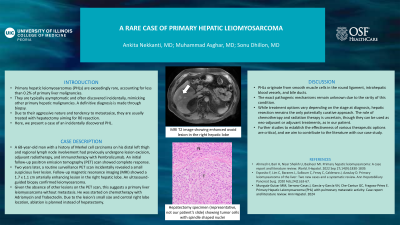
Introduction: Primary hepatic leiomyosarcomas (PHLs) are exceedingly rare, accounting for less than 0.2% of primary liver malignancies. They are typically asymptomatic and often discovered incidentally, mimicking other primary hepatic malignancies. A definitive diagnosis is made through biopsy. Due to their aggressive nature and tendency to metastasize, they are usually treated with hepatectomy aiming for R0 resection. Here, we present a case of an incidentally discovered PHL.
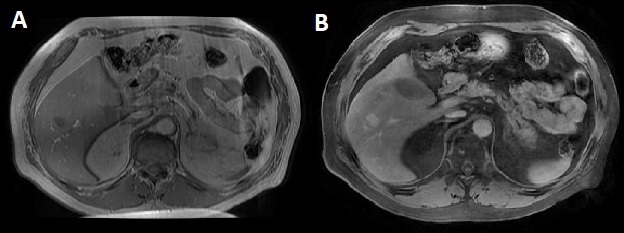
Case Description/Methods: A 68-year-old man with a history of Merkel cell carcinoma on his distal left thigh and regional lymph node involvement had previously undergone lesion excision, adjuvant radiotherapy, and immunotherapy with Pembrolizumab. An initial follow-up positron emission tomography (PET) scan showed complete response. Two years later, a routine surveillance PET scan incidentally revealed a small suspicious liver lesion. Follow-up magnetic resonance imaging (MRI) showed a 1.7 x 1.1 cm arterially enhancing lesion in the right hepatic lobe. An ultrasound-guided biopsy confirmed leiomyosarcoma. Given the absence of other lesions on the PET scan, this suggests a primary liver leiomyosarcoma without metastasis. He was started on chemotherapy with Adriamycin and Trabectedin. Due to the lesion's small size and central right lobe location, ablation is planned instead of hepatectomy. We anticipate this procedure and will monitor the patient's response to treatment.
Discussion: PHLs originate from smooth muscle cells in the round ligament, intrahepatic blood vessels, and bile ducts. The exact pathogenic mechanisms remain unknown due to the rarity of this condition. While treatment options vary depending on the stage at diagnosis, hepatic resection remains the only potentially curative approach. The role of chemotherapy and radiation therapy is uncertain, though they can be used as neo-adjuvant or adjuvant treatments, as in our patient. Further studies to establish the effectiveness of various therapeutic options are critical, and we aim to contribute to the literature with our case study.

Disclosures:
Ankita Nekkanti, MD, Muhammad Asghar, MD, Sonu Dhillon, MD. P1393 - Unexpected Diagnosis of Primary Hepatic Leiomyosarcoma, ACG 2024 Annual Scientific Meeting Abstracts. Philadelphia, PA: American College of Gastroenterology.
University of Illinois College of Medicine, Peoria, IL
Introduction: Primary hepatic leiomyosarcomas (PHLs) are exceedingly rare, accounting for less than 0.2% of primary liver malignancies. They are typically asymptomatic and often discovered incidentally, mimicking other primary hepatic malignancies. A definitive diagnosis is made through biopsy. Due to their aggressive nature and tendency to metastasize, they are usually treated with hepatectomy aiming for R0 resection. Here, we present a case of an incidentally discovered PHL.
Case Description/Methods: A 68-year-old man with a history of Merkel cell carcinoma on his distal left thigh and regional lymph node involvement had previously undergone lesion excision, adjuvant radiotherapy, and immunotherapy with Pembrolizumab. An initial follow-up positron emission tomography (PET) scan showed complete response. Two years later, a routine surveillance PET scan incidentally revealed a small suspicious liver lesion. Follow-up magnetic resonance imaging (MRI) showed a 1.7 x 1.1 cm arterially enhancing lesion in the right hepatic lobe. An ultrasound-guided biopsy confirmed leiomyosarcoma. Given the absence of other lesions on the PET scan, this suggests a primary liver leiomyosarcoma without metastasis. He was started on chemotherapy with Adriamycin and Trabectedin. Due to the lesion's small size and central right lobe location, ablation is planned instead of hepatectomy. We anticipate this procedure and will monitor the patient's response to treatment.
Discussion: PHLs originate from smooth muscle cells in the round ligament, intrahepatic blood vessels, and bile ducts. The exact pathogenic mechanisms remain unknown due to the rarity of this condition. While treatment options vary depending on the stage at diagnosis, hepatic resection remains the only potentially curative approach. The role of chemotherapy and radiation therapy is uncertain, though they can be used as neo-adjuvant or adjuvant treatments, as in our patient. Further studies to establish the effectiveness of various therapeutic options are critical, and we aim to contribute to the literature with our case study.

Figure: A. Pre-contrast T1 image showing hypointense ovoid lesion in the right hepatic lobe. B. Post-contrast T2 image showing enhancement of the lesion.
Disclosures:
Ankita Nekkanti indicated no relevant financial relationships.
Muhammad Asghar indicated no relevant financial relationships.
Sonu Dhillon indicated no relevant financial relationships.
Ankita Nekkanti, MD, Muhammad Asghar, MD, Sonu Dhillon, MD. P1393 - Unexpected Diagnosis of Primary Hepatic Leiomyosarcoma, ACG 2024 Annual Scientific Meeting Abstracts. Philadelphia, PA: American College of Gastroenterology.
