Sunday Poster Session
Category: Liver
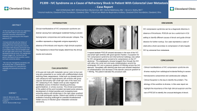
P1399 - IVC Syndrome as a Cause of Refractory Shock in Patient With Colorectal Liver Metastasis - Case Report
Sunday, October 27, 2024
3:30 PM - 7:00 PM ET
Location: Exhibit Hall E

Has Audio
- IM
Islam B Mohamed, MD
Cleveland Clinic Foundation
Cleveland, OH
Presenting Author(s)
Islam B Mohamed, MD1, Mohamad-Noor Abu-Hammour, MD2, Rashid Abdel-Razeq, MD3, Varun U. Shetty, MD1
1Cleveland Clinic Foundation, Cleveland, OH; 2Cleveland Clinic Fairview, Cleveland, OH; 3Cleveland Clinic, Cleveland, OH
Introduction: Clinical manifestations of IVC compression syndrome are diverse, varying from radiological incidental findings to severe hemodynamic compromise and cardiovascular collapse. The condition represents a diagnostic enigma, especially in the absence of thrombosis, and requires a high clinical suspicion. The impedance to blood flow largely determines the clinical course and outcome.
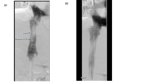
Case Description/Methods: A 53-year-old male with metastatic cancer colon to liver and lung who presented with shortness of breath, weakness and lightheadedness and was found to be in profound shock. Lab workup was significant for leukocytosis and high lactate. Point of care ultrasound (POCUS) showed a small right ventricle with hyper dynamic left ventricle with gross vascular abnormality with the small right side of the heart and IVC measuring 6 mm. He was initially treated with broad spectrum antibiotics due to presumed sepsis. Infectious work up was unrevealing for sources of infection. He developed clinical jaundice and increasing lower extremity edema with tenderness in the right upper quadrant and small peri-hepatic ascites which raise the suspicion of IVC syndrome. CT images showed ‘pancaking’ of the IVC by hepatic metastatic lesions, hepatomegaly, peri-hepatic ascites and paracaval lymphadenopathy. Conventional angiography showed sluggish flow through the IVC with reflux into the renal vein and collaterals around the intrahepatic IVC with a pressure gradient of 9 mmHg across the IVC stenosis. Venoplasty with stenting was done and showed resolution of collaterals and decrease of the stenotic pressure gradient to 1 MmHg. Unfortunately, he developed Takotsubo cardiomyopathy and acute liver failure. Considering his poor prognosis his health care POA opted to pursue comfort care and the patient passed away shortly after a companionate extubation.
Discussion: Obstruction of IVC by hepatic metastasis is a cause of IVC syndrome.The exact incidence of the disease in patients with metastatic liver deposits is unknown. Clinical presentation mimics hypo-volumic shock; however without high clinical suspicion the clinical syndrome can be a diagnostic dilemma especially in absence of IVC thrombosis. POCUS can be a useful tool especially in ICU setting to identify different causes of shock and guide clinical decision for further workup. Our case represents a case of Our case represents a case of obstructive shock secondary to compression of retro-hepatic IVC by colorectal liver metastasis causing transient Budd Chiari syndrome.

Disclosures:
Islam B Mohamed, MD1, Mohamad-Noor Abu-Hammour, MD2, Rashid Abdel-Razeq, MD3, Varun U. Shetty, MD1. P1399 - IVC Syndrome as a Cause of Refractory Shock in Patient With Colorectal Liver Metastasis - Case Report, ACG 2024 Annual Scientific Meeting Abstracts. Philadelphia, PA: American College of Gastroenterology.
1Cleveland Clinic Foundation, Cleveland, OH; 2Cleveland Clinic Fairview, Cleveland, OH; 3Cleveland Clinic, Cleveland, OH
Introduction: Clinical manifestations of IVC compression syndrome are diverse, varying from radiological incidental findings to severe hemodynamic compromise and cardiovascular collapse. The condition represents a diagnostic enigma, especially in the absence of thrombosis, and requires a high clinical suspicion. The impedance to blood flow largely determines the clinical course and outcome.
Case Description/Methods: A 53-year-old male with metastatic cancer colon to liver and lung who presented with shortness of breath, weakness and lightheadedness and was found to be in profound shock. Lab workup was significant for leukocytosis and high lactate. Point of care ultrasound (POCUS) showed a small right ventricle with hyper dynamic left ventricle with gross vascular abnormality with the small right side of the heart and IVC measuring 6 mm. He was initially treated with broad spectrum antibiotics due to presumed sepsis. Infectious work up was unrevealing for sources of infection. He developed clinical jaundice and increasing lower extremity edema with tenderness in the right upper quadrant and small peri-hepatic ascites which raise the suspicion of IVC syndrome. CT images showed ‘pancaking’ of the IVC by hepatic metastatic lesions, hepatomegaly, peri-hepatic ascites and paracaval lymphadenopathy. Conventional angiography showed sluggish flow through the IVC with reflux into the renal vein and collaterals around the intrahepatic IVC with a pressure gradient of 9 mmHg across the IVC stenosis. Venoplasty with stenting was done and showed resolution of collaterals and decrease of the stenotic pressure gradient to 1 MmHg. Unfortunately, he developed Takotsubo cardiomyopathy and acute liver failure. Considering his poor prognosis his health care POA opted to pursue comfort care and the patient passed away shortly after a companionate extubation.
Discussion: Obstruction of IVC by hepatic metastasis is a cause of IVC syndrome.The exact incidence of the disease in patients with metastatic liver deposits is unknown. Clinical presentation mimics hypo-volumic shock; however without high clinical suspicion the clinical syndrome can be a diagnostic dilemma especially in absence of IVC thrombosis. POCUS can be a useful tool especially in ICU setting to identify different causes of shock and guide clinical decision for further workup. Our case represents a case of Our case represents a case of obstructive shock secondary to compression of retro-hepatic IVC by colorectal liver metastasis causing transient Budd Chiari syndrome.

Figure: Figure B: Digital subtraction angiography pictures
B1: Showing retro-hepatic IVC with stenosis with collaterals (arrow)
B2: restoration of blood flow and resolution of collaterals
B1: Showing retro-hepatic IVC with stenosis with collaterals (arrow)
B2: restoration of blood flow and resolution of collaterals
Disclosures:
Islam B Mohamed indicated no relevant financial relationships.
Mohamad-Noor Abu-Hammour indicated no relevant financial relationships.
Rashid Abdel-Razeq indicated no relevant financial relationships.
Varun Shetty indicated no relevant financial relationships.
Islam B Mohamed, MD1, Mohamad-Noor Abu-Hammour, MD2, Rashid Abdel-Razeq, MD3, Varun U. Shetty, MD1. P1399 - IVC Syndrome as a Cause of Refractory Shock in Patient With Colorectal Liver Metastasis - Case Report, ACG 2024 Annual Scientific Meeting Abstracts. Philadelphia, PA: American College of Gastroenterology.

